Keywords
Evaluation Studies as Topic; Islets of Langerhans; Rats
Abbreviations
IUR: isotropic uniform random
INTRODUCTION
The mean volume of the islets and the total number, as well as the volume, of the beta cells in human or animal pancreas are fundamental parameters under developmental, aging, pathological, and therapeutic conditions. Reduced islet number and/or diminished volume of the beta cells mass in the pancreas of diabetic subjects have been reported by many authors [1]. Using stereological methods, qualitative variables, including hyperplasia/hypoplasia and hypertrophy/ atrophy, can be defined through quantitative and comparable data. As a result, the impact of different conditions on the beta cell survival or changes needs simple and rapid methods of quantification.
The pioneers of stereology have described the unbiased methods of quantification, including the estimation of the number as well as the volume of the beta cells [2, 3]. Recently, pancreas of mice have been examined by using state-of-the-art quantitative-stereological methods in order to obtain the total islet volume, beta cell volume, and the absolute number of the beta cells [4]. Other investigators have examined the nuclei counting for measuring the total number of the cells in islet preparations after disrupting the cells and the islets with lysis solution and shearing. All of these methods are valuable; however, there are some limitations, including serial sectioning of the tissues. Also, using a physical disectors for cell counting may take a long time to prepare.
The present study aims to describe a stereological method which does not need the serial sectioning. On the other hand, it requires only a limited number (i.e., 10-13) of the isotropic uniform random slabs of each pancreas to be embedded in a paraffin block. Moreover it needs one 5 μm and one 20 μm micrometer section to be cut using a microtome and each one to be mounted on a microscopic slide in order to estimate the volume of the pancreas, the total volume of the islets, and the total number, as well as the mean volume, of the beta cells.
METHODS
Animal Model
Five male Sprague-Dawley rats weighing between 210 and 290 g were selected. They were housed in the laboratory with standard conditions including 12 h dark/light cycle in rooms with a controlled temperature (22-26°C) as well as free access to food and water.
Pancreas Volume and Shrinkage Estimation
The pancreas was removed and cleaned of fatty and connective tissues. Then, the pancreas was weighed and the primary volume V(primary) of the pancreas was measured using the immersion method [5]. The surrounding fat was trimmed away carefully such that measured volume actually reflected the volume of the pancreas. As some tissue deformation could occur in the form of shrinkage produced by fixation, tissue processing, and staining, which will influence the stereological estimation, the shrinkage had to be estimated. The shrinkage was also used for estimating of the final pancreas volume after tissue processing without the need for serial sectioning, which is required for the “Cavalieri method”. Estimation of shrinkage and cell volume (using local estimators), length or surface area requires sections that are isotropic in three dimensions, (i.e., they do not have a preferred orientation requires isotropic uniform random (IUR) sectioning of the pancreas [6, 7, 8, 9]. IUR sectioning means that the direction and position of cutting could not have been predicted in advance, thus, it is random in 3D. It should be noted that the shrinkage factors not only require isotropic uniform random sectioning but they also assume that shrinkage is concentric, i.e. it affects all components of the pancreas in the same way. These sections were prepared by the “orientator method” [10]. An explanation of IUR sectioning has been given as the legend of Figure 1. Briefly, the pancreas was placed at the center of a circle with equal divisions. A random number was selected and the tissue was sectioned. Each piece of the sectioned tissue was placed on a second circle with non-equal divisions (the divisions were cosine-weighted). The piece of the tissue was reoriented such that the tissue was resting on the new cut surface and the original base now was flush with the 0-0 line of the second circle. Another random number was selected and the tissue was sectioned into slabs. Then, another piece of the tissue was placed vertically on the same circle and sectioned into slabs in a new direction. These slabs were cut with constant thickness and a random start. On the average, 10-13 slabs from each pancreas were obtained. All the slabs were collected. The second cut surfaces of the slabs are the IUR surfaces. Using a trocar, two circular pieces (about 1 mm diameter) were punched from the IUR surfaces of the two slabs were randomly selected. The diameter and the area of the circular pieces of the pancreas were measured. For fixation, the pancreas was placed in neutral buffered formaldehyde and afterwards all the collected slabs and the circular pieces were embedded in the same paraffin block. One 5 μm and one 20 μm sections were cut using a microtome. Then, each one was mounted on a microscopic slide. The 5 μm section was used in order to estimate the volume of the islets as well as the mean volume of the beta cells, and the 20 μm section was used in order to obtain the total number.
Figure 1. Orientator method to obtain isotropic uniform random
(IUR) sections. a. The pancreas was placed at the center of a circle
with equal divisions. A random number was selected and the tissue
was sectioned (here 2). Each part of the sectioned tissue was placed
on a second circle with non-equal divisions. b. Another random
number was selected (here 6) and the tissue was sectioned into slabs. c. Another part was vertically placed on the same circle and
sectioned into slabs in a new direction (here 4). d. All the slabs were
sampled. Using a trocar, two circular pieces (about 1 mm diameter)
were punched randomly from pancreas IUR surfaces of the slabs. e. All the slabs and the circular pieces were embedded in the same
block of paraffin (the arrows). f. After staining of the sections, the
area of the circular pieces was measured again.
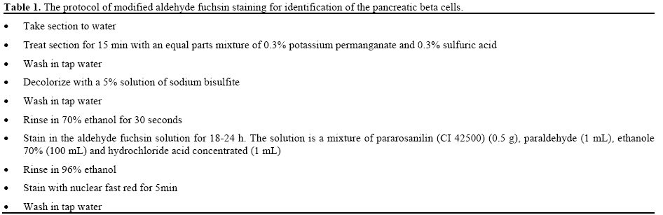
After tissue sectioning, processing, and staining of the sections, the area of the circular pieces was measured again (Figure 1). The tissue sections stained with modified aldehyde fuchsin according to Bangler [11]. Due to substantial modification, a description of the staining is presented in Table 1.
The degree of shrinkage d(sh) was calculated using the following formula:
d(sh) = 1- ( AA / AB ) 1.5
where AA and AB are the area of each circular piece of the pancreas after and before processing, sectioning, and staining, respectively.
Volume Density of the Pancreatic Islets
Each microscopic slide was analyzed using a video microscopy system which was made up of a microscope (E-200, Nikon, Tokyo, Japan) linked to a video camera, a computer, a flat monitor, and a microcator (MT-12, Heidenhain, Traunreut, Germany). By means of the stereology software designed at our laboratory (Stereological Research Laboratory, Shiraz University of Medical Sciences, Shiraz, Iran), the stereological probes (composed of test points and unbiased counting frame) were superimposed upon the tissue images viewed on the monitor.
Moreover, the fields of each microscopic slide were sampled in a systematic random manner and analyzed using a microscope at the final magnification of 340x (Figure 2). The sampling was randomly started from a corner of the slide out of the tissue sections. Then, the slide was moved in the X- and Y-directions at equal distances. The point counting method was used to estimate the volume density of the islet on 5 μm thickness sections (Figure 2) [6, 7, 8, 9]. The average number of the points hitting the reference space (the pancreas) and the islets ranged 260-300 and 2-4, respectively. The volume density (Vv) of the pancreatic islet was estimated using the following formula:
Vv = P(islet) / P(reference)
Figure 2. a. Systematic random sampling of the microscopic fields. b. Volume density estimation of the pancreatic islets (the
arrowheads) using the point counting method. The arrow indicates
the point which exists on the right upper corner of the cross.
where P(islet) and P(reference) were the number of the test points falling on the islet’s profile and on the reference space, respectively. The following formula was used to estimate the final islet volume:
V(islets) = Vv x V(primary) x (1 - d(sh)).
By multiplying the volume density of the islet by the final volume of the pancreas, the absolute volume of the parameters was estimated to prevent the “reference trap” [12, 13]. The term “reference trap” refers to the fact that interpretations solely based on densities may lead to wrong conclusion.
Numerical Density of the Beta Cell
The numerical density of the beta cells was estimated on 20 μm thickness. An oil immersion objective lens (100x) with the numerical aperture of 1.4 was used at the final magnification of 3,400x in order to count the cells using the optical disector method (Figure 3) [6, 7, 8, 9].
Figure 3. Optical scan for estimation of the number of the beta cells.
Just the cells that whose nuclei did not appear in the beginning of the
disector height (a.) and appeared at the following optical scan of the
disector height (b.) were counted. The cells whose nuclei were
completely or partly inside the counting frame or touched the upper
and right lines were considered as "ΣQ
_" (the arrows). c. A schematic
drawing of a microscopic slide with a thick section of pancreas
mounted on it. t is the mean final section thickness measured using
the microcator. h is the height of the optical disector. In each field,
upper and lower guard zones (gz) were ignored. The counting frames
were overlaid on the entire areas of the islets. d. Point-sampled
intercept method. The cells were sampled using a point grid and the
volume was estimated by measuring the length of the intercept. Here
the intercepts of 3 beta cells are shown.
The standard approach of a systematic grid of frames in the pancreas would lead to about 99% of the frames hitting the tissue outside the islands. Therefore, an average of about 110 counting frames was applied per pancreatic islands. The slide of pancreas was looked for the islands at a low magnification. Since there was a limited number of the islands found in the sections of each pancreas, all the entire island which were observable in low magnification were considered for evaluating at high magnification. As previously mentioned the sections of the pancreas were obtained using IUR sectioning. Therefore, the evaluated islands were randomly selected. Then, the whole area of each island was considered for counting. In each field, the first 5 μm of the section thickness was ignored to avoid biased counting (guard zone). Cell counting was done in the next 10 μm of the section thickness which has been called “height of disector” or “h”. Just the cells whose nuclei did not appear in the beginning of the disector height and appeared at the following optical scan of the disector height were counted. The cells whose nuclei were completely or partly inside the counting frame or touched the upper and right lines were counted (ΣQ _) (Figure 3). The numerical density (NV) was estimated using the optical disector method and the following formula:
NV = ( ΣQ _ / ( h x a/f x Σp) ) x ( t / BA)
where, a/f is the area of the counting frame (here 184 μm2), h is the height of the optical disector (here 10 μm), ΣQ _ is the number of the beta cells counted in all the disectors, Σp is the total number of the counted frames, BA is the microtome block advance to cut the block (here was 20 μm), and t is the mean of the final section thickness (17.4 μm on the average). In addition, t was measured using a microcator on a random field of each islet and mean value was calculated [14]. On the average 9 islets were recognizable in each microscopic slide.
To estimate the total number of the beta cells (N(beta cell)) the following formula was used:
N(beta cell) = NV x V(islets)
where V(islets) is the total islet volume
Volume of the Beta Cell
Point-sampled intercept method was used to obtain the volume-weighted mean volume of the cells [9]. The mean cell volume (Vv) was estimated on the sampled cells using a point grid. In this method, first, the cells were sampled according to their volume using the point on the thinner section (5 μm). On the second step, the volume was estimated by measuring the length of the intercept. The intercept is a line which passes through the point hitting the cells and extends to the cell borders (Figure 3). Also, due to isotropic uniform random sectioning of the pancreas, the direction of the measurement was selected horizontally.
The intercept length (l0) was measured from the appeared nuclei to the right and left side of the cell border in the horizontal axis. The volume density was estimated using the following formula:
Vv = π / 3 x mean( l0 3 )
ETHICS
The animals were treated according to the standard directive as recommended and approved by the research authorities of Shiraz University of Medical Sciences (Laboratory Animal Ethics Committee).
STATISTICS
Data were expressed as mean±SD and coefficient of variation (CV).
RESULTS
On average, 10-13 slabs, one 5 μm, and one 20 μm micrometer sections were collected per rat pancreas. On the average, 200 cells were counted in each pancreas.
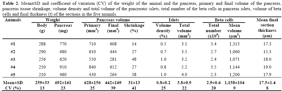
Table 2 shows the results obtained from the five rats. After calculation of the tissue shrinkage (about 30% on the average), the primary volume (628±156 mm3) was corrected in order to obtain the final volume (442±169 mm3). The real thickness of the sections on the microscopic slides was 11.7 μm. Moreover, the mean islet volume was 3.8±0.9 mm3 and the total number of the beta cells was 2.9x10⁶. Also, the volume-weighted mean-volume of the beta cells was measured as 1,158±104 μm3. It takes one hour to estimate the volume of the islets and two hours to count the cells and estimate the intercepts per animal.
DISCUSSION
The present study described a stereological method for estimating the total number of the beta cells and the mean beta cell volume in the rat pancreas through light microscopy. Simplicity of counting and fewer sections was the first part of our goal. In the study conducted in 1999 by Bock et al., exhaustive sections of pancreas were prepared based on the fractionator principle [3]. Their work is an unbiased estimator of the number, as well as the volume, of the beta cells. Nevertheless, exhaustive sectioning is time-consuming. In addition, a physical disector was used in that study (i.e., using two physical sections with a determined distance). Finding cells according to the method is also time-consuming. On the other hand, the present study relies on shrinkage correction of the tissue and sections. It takes almost one hour to estimate the volume of the islets and two hours to count the cells and estimate the intercepts per animal. Also, since tissue shrinkage of blocks of paraffin is more than plastic blocks, the tissue shrinkage should be corrected after embedding and sectioning. Although we suggested a reasonable approach for correcting the global shrinkage, it should be noted that the pancreatic islets might not be shrunk the same as the surrounding pancreatic tissue. However, there is no method to estimate the shrinkage of the islets for which no corrections can be calculated from the generated sections. One of the important advantages of the study by Bock et al. is that “fractionator method” is independent of shrinkage [3]. The method by Bock et al. needs exhaustive sectioning and it is time-consuming.
Another part of our goal was cost-beneficial staining of the beta cells and applying an “optical disector”. Although immunohistochemical stain for insulin makes an easier approach to cell identification, aldehyde fuchsin was used here. A limitation of this type of staining might be confusion in identification of the cells and their borders in thick sections. The thick section is recommended for counting by “optical disector method”. In routine staining methods the cells cannot be recognized well. The Table 1 presents the modification of the staining. Nevertheless, the method can be improved using the immunohistochemical stain for insulin, plastic embedding, and thicker sections. However, it should be remembered that it is difficult to penetrate an immunostain into the thick plastic sections. As investigators have recommended, the volume density of the islets was estimated on 5 μm thick sections in order to prevent overprojection. As a general rule, overprojection is negligible if the section thickness is less than 10% of the diameter of the particle (the islets). Mac Gregor et al. in 2006 reported that most small islets have a diameter of 75-100 μm, whereas large islets have a diameter of about 200 μm [15].
Bock et al. reported about 2.7x10⁶ beta cells per pancreas with a mean cell volume of 1,170 μm3 in 47- day-old male Lewis/MOL rats [3]. Chintinne et al. in 2010 showed that pancreas of three Wistar rats contained 2.8±0.2x10⁶ beta cells [16]. Moreover, Herbach et al. in 2011 reported about 1.3x10⁶ beta cells per pancreas with the mean cell volume of about 1,300 μm3 in female Charles River mice [4]. In another study published by Herbach et al. in 2005, no significant difference was found regarding the development of the total islet and beta cell volumes between male and female control mice [17]. Sato and Herman in 1981 also reported a value of the fractional volume of the islets of about 2.2% and an average beta cell volume of 1,260 μm3 in the rabbit pancreas [2]. Our findings are also in accordance with these studies.
The volume density was estimated using the point counting routine with one grid of points. To spending less time counting hits over the total pancreas area it is recommended to use a multipurpose grid, i.e. to have two grids, one for the reference area and another for the beta cells. These two grids usually are combined.
Morphometrical study of the pancreas has also been reported by the investigators of this field. For instance, Adeyemi et al. in 2010 reported the number of the beta cells per islet as 10.64/1,000 μm2 with a mean cell volume of 115 μm3 in Wistar rats [18]. As the pioneer stereologists have stated, this type of study could be biased and obtaining the data using the unbiased stereological methods improves the quantitative study.
CONCLUSIONS
The present study described a simple method for stereological estimation of the volume of the pancreas, the volume of the islets, and the total number, as well as the mean volume, of the beta cells by using a limited number of the sections of pancreas mounted on a microscopic slide. It takes one hour to estimate the volume of the islets and two hours to count the cells and estimate the intercepts per animal.
Acknowledgement
The present work was financially supported by Histomorphometry and Stereology Research Center, Shiraz University of Medical Sciences, Shiraz, Iran. Research Improvement Center of Shiraz University of Medical Sciences and Ms. Afsaneh Keivanshekouh are also appreciated for improving the use of English in the manuscript
Conflict of interest
The authors report no conflicts of interest
References
- Marchetti P, Lupi R, Del Guerra S, Bugliani M, Marselli L, Boggi U. The beta-cell in human type 2 diabetes. Adv Exp Med Biol. 2010;654:501-14. [PMID 20217512].
- Sato T, Herman L. Stereological analysis of normal rabbit pancreatic islets. Am J Anat. 1981;161(1):71-84. [PMID 7018211]
- Bock T, Svenstrup K, Pakkenberg B, Buschard K. Unbiased estimation of total beta-cell number and mean beta-cell volume in rodent pancreas. APMIS. 1999;107(8):791-9. [PMID 10515130]
- Herbach N, Bergmayr M, Göke B, Wolf E, Wanke R. Postnatal development of numbers and mean sizes of pancreatic islets and beta-cells in healthy mice and GIPR(dn) transgenic diabetic mice. PLoS One. 2011;6(7):e22814. [PMID 21818396]
- Sherle W. A simple method for volumetry of organs in quantitative stereology. Mikroskopie. 1970; 26(1): 57-60.
- Boyce RW, Dorph-Petersen KA, Lyck L, Gundersen HJ. Design-based stereology: introduction to basic concepts and practical approaches for estimation of cell number. Toxicol Pathol. 2010;38(7):1011-25. [PMID 21030683]
- Nyengaard JR. Stereologic methods and their application in kidney research. J Am Soc Nephrol. 1999;10(5):1100-23. [PMID 10232698].
- Gundersen HJ, Bendtsen TF, Korbo L, Marcussen N, Møller A, Nielsen K, Nyengaard JR, Pakkenberg B, Sørensen FB, Vesterby A, et al. Some new, simple and efficient stereological methods and their use in pathological research and diagnosis. APMIS. 1988;96(5):379- 94. [PMID 3288247 ]
- Gundersen HJ, Bagger P, Bendtsen TF, Evans SM, Korbo L, Marcussen N, Møller A, Nielsen K, Nyengaard JR, Pakkenberg B, et al. The new stereological tools: disector, fractionator, nucleator and point sampled intercepts and their use in pathological research and diagnosis. APMIS. 1988;96(10):857-81. [PMID 3056461].
- Mattfeldt T, Mall G, Gharehbaghi H, Möller P. Estimation of surface area and length with the orientator. J Microsc. 1990;159(Pt 3):301-17. [PMID 2243364].
- Bangler Jr. Factors influencing the staining of beta-cell granules in pancreatic islets with various basic dyes, including paraldehydefuchsin. Am J Pathol. 1956;32(2):349-62. [PMID 13302402].
- Braendgaard H, Gundersen HJ. The impact of recent stereological advances on quantitative studies of the nervous system. J Neurosci Methods. 1986;18(1-2):39-78. [PMID 3540470]
- Gruber C, Kohlstedt K, Loot AE, Fleming I, Kummer W, Mühlfeld C. Stereological characterization of left ventricular cardiomyocytes, capillaries, and innervation in the nondiabetic, obese mouse. Cardiovasc Pathol. 2011: 22. [PMID 22197049]. Epubhead of print.
- Dorph-Petersen KA, Nyengaard JR, Gundersen HJ. Tissue shrinkage and unbiased stereological estimation of particle number and size. J Microsc. 2001;204(Pt 3):232-46. [PMID 11903800]
- MacGregor RR, Williams SJ, Tong PY, Kover K, Moore WV, Stehno-Bittel L. Small rat islets are superior to large islets in in vitro function and in transplantation outcomes. Am J Physiol Endocrinol Metab. 2006;290(5):E771-9. [PMID 16303846].
- Chintinne M, Stangé G, Denys B, In 't Veld P, Hellemans K, Pipeleers-Marichal M, Ling Z, Pipeleers D. Contribution of postnatally formed small beta cell aggregates to functional beta cell mass in adult rat pancreas. Diabetologia. 2010;53(11):2380-8. [PMID 20645074]
- Herbach N, Goeke B, Schneider M, Hermanns W, Wolf E, Wanke R. Overexpression of a dominant negative GIP receptor in transgenic mice results in disturbed postnatal pancreatic islet and beta-cell development. Regul Pept. 2005; 125(1-3):103-17. [PMID 15582721]
- Adeyemi DO, Komolafe OA, Adewole OS, Obuotor EM, Abiodun AA, Adenowo TK. Istomorphological and morphometric studies of the pancreatic islet cells of diabetic rats treated with extracts of Annonamuricata. Folia Morphol (Warsz). 2010;69(2):92- 100.


