Keywords
Immunoglobulin G; Pancreatic Neoplasms; Pancreatitis, Chronic
INTRODUCTION
Autoimmune pancreatitis is a disorder which predominantly affects the pancreas. Over the past few years this condition has been extensively studied and there is an increasing consensus that autoimmune pancreatitis is in fact a multisystem disorder predominantly affecting the pancreas. Most patients respond to steroids but occasionally additional immunosuppression may be required.
We report a case of pericardial involvement in autoimmune pancreatitis. The case is also complicated by the fact that he did not respond to steroids and required 6-mercaptopurine for control of his disease.
CASE REPORT
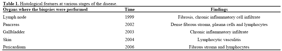
A sixty-year-old gentleman was admitted under the surgical team with a two week history of abdominal pain and dark urine in October 2001. There was no history of jaundice, itching, weight loss or loss of appetite. He suffered from left leg lymphedema, Raynauds disease, transient ischemic attacks, ischemic heart disease and had left sided hydronephrosis. He is a chronic smoker but does not consume any alcohol. He had lymph node biopsy performed in 1999. There was no history of risk factors for liver disease. The positive findings on examination were jaundice and lymphedema of his left leg. The liver function tests revealed a cholestatic pattern: total bilirubin 5.15 mg/dL (reference range: 0-1.11 mg/dL), alanine transaminase 96 IU/L (reference range: 0-40 IU/L), alkaline phosphatase 387 IU/L (reference range: 35- 120 IU/L) and gamma-glutamyl transpeptidase 278 IU/L (reference range: 0-50 IU/L). Transabdominal ultrasound revealed generalised biliary dilatation. The patient underwent an ERCP, which revealed a lower common bile duct stricture. A plastic biliary stent was inserted for drainage. The CA 19-9 was 126 U/mL (reference range: 0-30 U/mL). The patient had a computed tomography (CT) scan (Figure 1) performed, which revealed a 3 cm mass in the head of the pancreas. Radial endoscope ultrasound confirmed the CT finding but also showed that the mass was encasing the superior mesenteric vein. In view of these findings a diagnosis of pancreatic cancer was considered. A laparoscopy guided biopsy of the mass was performed in March 2002 and this revealed diffuse replacement of the pancreatic parenchyma by a fibrous stroma with plasma cell infiltration and storiform fibrosis (Figure 2). The patient continued to be jaundiced and the tumour marker CA 19-9 increased to 3,265 U/mL. The decision was made to perform a palliative bypass surgery for relief of symptoms. The operation confirmed the CT and the EUS findings and therefore the patient underwent a gastrojejunostomy, choledochojejunostomy and a cholecystectomy in 2002. All the biopsies (pancreas, gallbladder, liver and the lymph node) were reviewed by the specialist histopathologist and a diagnosis of sclerosing retroperitonitis was raised (Table 1). Meanwhile the patient developed a nodular itchy rash during the intervening period. The skin biopsies confirmed similar findings. At this point his case was reviewed and a diagnosis of autoimmune pancreatitis postulated. Total immunoglobulin G (IgG) and subclass assay was performed; results were as follows: IgG1, 17.84 g/L (reference range: 2.2-10.4 g/L); IgG4: 9.48 g/L (reference range: 2.0-2.4 g/L); IgG2 and IgG3 were within the normal range. Previous pancreatic histology was reviewed and was consistent with a diagnosis of autoimmune pancreatitis.
Figure 1. CT scan showing 3 cm mass (arrow) in the head of
pancreas.
Figure 2. Pancreatic core biopsy showing lymphoplasmacytic
sclerosing fibrosis (a.) and storiform fibrosis (b.).
The patient was started on prednisolone 40 mg od. He responded very well to treatment but his symptoms relapsed when the dose was tapered. He was restarted on the same dose of prednisolone, i.e. 40 mg/day. Repeat CT scan showed good resolution of the mass. During this period he was admitted with signs of right heart failure. He was on prednisolone 30 mg/day as the treatment dose in this admission. The liver function tests had worsened and the immunoglobulin G levels were elevated .The echocardiogram was normal but a magnetic resonance scan of the heart revealed pericardial thickening (Figure 3). The patient underwent a pericardectomy. Intraoperative findings revealed a thickened pericardium and the surgeon performed pericardial biopsies. The histopathology findings revealed fibrous stroma and lymphocytes and stained strongly for IgG4 (Figure 4). As he had relapsed twice whilst on steroids we started him on azathioprine. Unfortunately he did not respond/tolerate azathioprine and he was started on 6-mercaptopurine. Over a period of time the immunoglobulin levels and liver function tests have gradually improved. His steroid dose was tapered at the rate of 5 mg/week and is now maintained on prednisolone 10 mg/day and 6- mercaptopurine 75 mg/day.
Figure 3. MR scan showing pericardial thickening.
Figure 4. Pericardial biopsy with a strongly positive IgG4 stain.
DISCUSSION
Autoimmune pancreatitis has been described as a primary pancreatic disorder and also in association with other diseases of presumed autoimmune aetiology including sclerosing cholangitis, primary biliary cirrhosis, retroperitoneal fibrosis, rheumatoid arthritis, sarcoidosis, and Sjögren’s syndrome [1]. The diagnosis of autoimmune pancreatitis is based on a combination of clinical, radiological, serological and histological findings [2]. The mean age of presentation is in the sixth decade with the age range between 30 to 80 years. They classical present with a pancreatic mass in the head of the pancreas [3] as in our patient. They may also present with jaundice, abdominal pain, weight loss and diabetes mellitus. In confirmed cases of autoimmune pancreatitis effort should be made to look for other systemic extra pancreatic manifestations of the disease. Imaging is required to arrive at the diagnosis.
Immunology of Autoimmune Pancreatitis
Thought the exact cause of autoimmune pancreatitis is uncertain immunological mechanisms play a vital role. This theory is consolidated by many findings including prevalence of hypergammaglobulinemia, auto antibodies, specific immune mediated histological changes and response to steroids. The immunoglobulin subclass, IgG4, has found to be significantly raised in this condition as compared to other autoimmune disorders and a positive stain can be found in up to 85 % of the tissues [4]. The trigger for the IgG4 elevation and its pathogenetic role in autoimmune pancreatitis are unknown but one should interpret this with caution as IgG4 can be elevated in certain malignancies. The association of autoimmune pancreatitis with other similar conditions, i.e. retroperitoneal fibrosis, has resulted in the hypothesis that this could indeed be an IgG4 related sclerosing process.
Though autoimmune pancreatitis should be considered in all patients with a pancreatic mass it still remains a rare disorder. The clinician should have a low threshold for diagnosis as early treatment results in spontaneous resolution of signs and symptoms.
As mentioned earlier when encountering a case of autoimmune pancreatitis with elevated levels of gammaglobulins, IgG, and IgG4, an effort should be made to detect other systemic extrapancreatic abnormalities. There is evidence that early administration of steroid therapy is helpful in patients with autoimmune pancreatitis and retroperitoneal fibrosis. Our case is unusually complex as there are a number of organs (i.e. pancreas, lymph node, liver, gallbladder, retroperitoneum, skin and pericardium) were involved at different times of the disease process. Moreover, this is the second reported case of pericardial involvement in autoimmune pancreatitis in literature. Sugimoto et al. reported a case describing constrictive pericarditis as an initial manifestation of hyper-IgG4 disease [5].
The overall prognosis is said to be good as the disease is self limiting in majority of the cases with or without long term pancreatic dysfunction. There have been numerous reports on dramatic response to steroids but spontaneous response is also seen without treatment [6]. Some patients may require low dose maintenance therapy.
There is very little data in the literature regarding use of other immunosuppresives [7]. Response to steroids in our patient was not sustained on discontinuation necessitating the use of azathioprine and 6- mercaptopurine.
In conclusion, this case is unique because of the involvement of a number of organs in a single individual, i.e. lymph node, liver, pancreas, gall bladder, skin and the pericardium. A selected group of patients will have extensive multisystem disease that may not respond to steroids and early immunosuppression with alternative agents should be considered. This case also reinforces the observations by various authors that autoimmune pancreatitis is probably an IgG4 related diffuse autoimmune disease.
Acknowledgement
`All the authors have been actively involved in the management of the patient and the preparation of the manuscript
Conflict of interest There are no conflicts of interests to report
References
- Kamisawa T, Egawa N, Nakajima H. Autoimmune is a systemic autoimmune disease. Am J Gastroenterol. 2003 ;98(12):2811-2
- Yoshida K, Toki e, Takeuchi T et al. chronic pancreatitis caused by an autoimmune abnormality: proposal of the concept of autoimmune pancreatitis. Dig Dis Sci 1995; 40:1561-68
- Wakabayashi T, Kawaura Y, Satomura Y et al. Clinical and imaging features of autoimmune pancreatitis with focal pancreatic swelling or mass formation: comparison with so-called tumor-forming pancreatitis and ancreatic carcinoma. Am J Gastroenterol. 2003 ;98(12):2679-87
- Deheragoda MG, Church NI, Rodriguez - Justo M et al. The use of immunoglobulin Ig4 immunostaining in diagnosing pancreatic and extrapancreatic involvement in autoimmune pancreatitis. Clin Gastroenterol patol. 2007 Oct;5(10):1229-34.
- Constrictive pericarditis as an emerging manifestation of hyper-IgG4 disease. Sugimoto T, Morita Y, Isshiki K et al. Int J Cardiol. 2008 Nov 28;130(3):e100-1.
- Pearson RK, Longnecker DS, Chari ST et al. Controversies in linical pancreatology: autoimmune pancreatitis: does it exist? Pancreas. 2003 ;27(1):1-13 7.
- Church NI, Pereira SP, Deheragoda MG et al. Autoimmune pancreatitis: clinical and radiological features and objective response to steroid therapy in a UK series. Am J Gastroenterol. 2007 Nov;102(11):2417-25.


