Research Article - (2023) Volume 7, Issue 1
Biosynthesis of Silver Nanoparticles using Drimia indica and Exploring its Antibacterial Profile
Pratik S. Kamble,
Jayashree P. Gadade,
Dhanashree S. Patil,
Mamata A. Jagtap,
Dayanand P. Jayannawar,
Mansingraj S. Nimbalkar and
Swaroopa A. Patil*
Department of Botany, Shivaji University, Kolhapur, India
*Correspondence:
Swaroopa A. Patil,
Department of Botany, Shivaji University, Kolhapur,
India,
Email:
Received: 02-Jan-2023, Manuscript No. IPNNR-22-15313;
Editor assigned: 04-Jan-2023, Pre QC No. IPNNR-22-15313 (PQ);
Reviewed: 18-Jan-2023, QC No. IPNNR-22-15313;
Revised: 23-Jan-2023, Manuscript No. IPNNR-22-15313 (R);
Published:
30-Jan-2023, DOI: 10.12769/ipnnr-23.7.01
Abstract
Biological synthesis reflects as an eco-friendly, nontoxic and easy method of nanoparticle preparation. Present investigation
deals with biosynthesis of silver nanoparticles using Drimia indica leaf extract. Initially the synthesized
silver nanoparticles were characterized and confirmed by UV-Vis spectroscopy, X-ray diffraction (XRD) and SEM. The
synthesized nanoparticles when used for determination of antibacterial activity, by Microtitre Broth Dilution method
exhibited remarkable activity against Pseudomonas aeruginosa, Klebsiella pneumoniae and Escherichia coli.
Keywords
Drimia indica, Klebsiella, Pseudomonas, Escherichia, Microtitre Broth Dilution
Introduction
Nanotechnology is the most immerging branch of Science and
Technology. It is one of the most promising technologies applied
in all areas of Science. Apart from its tremendous applications
in Pharmaceuticals, textiles industries, electronics etc,
it is ecologically sound and cost effective. Metal nanoparticles
have received global attention due to their enormous applications
in the biomedical and physiochemical fields. Nanoparticles
aqueduct the space between bulky materials and molecular
structures [1]. Synthesizing metal nanoparticles using plants
has been recognized as green and efficient way for further exploiting
plants as convenient nanofactories [2]. Nanoparticles
with antimicrobial activity are advantageous in reducing acute
toxicity, lowering cost and overcoming resistance as compared
to other prevalent antibiotics [3,4].
Antibiotics are used as antimicrobial agents in medical field.
Higher doses of antibiotics causes harm to human cells and
forms cancerous cells and mutations. Use of nanoparticles as
antimicrobial agents is increasing day by day. The over doses
of antibiotics harms human cells, causes paralysis with more
disabilities.
Continued use of antibiotics for treatment of an array of inContinued use of antibiotics for treatment of an array of infections
has imposed the danger of developing antibiotic resistance.
Widespread bacterial infection treatment regime
involves higher initial doses of antibiotics followed by gradual
lowering down the doses, which requires a longer span of time.
A scenario of increased initial doses is practiced regularly which
has developed antibiotic resistance for a specific microorganism
or a set of microorganisms. Treatment plans for dreadful
infections having selective antibiotics for use are compromised
due to development of antibiotic resistance, as single antibiotic
is used for treatment of such infections. Control measures
may be possible in case of broad spectrum antibiotic used or
in organisms that are non-selective to antibiotics. On the other
hand there are infections which need specific/selective antibiotics
for cure. If resistance for single known antibiotic is developed
in organisms, the treatment lines may go unattended or
may fail totally. There is a need of promising interventions of
multifaceted alternative products to antibiotics which can play
a vital role in the field of medical science.
Biogenic nanoparticles are used as replacement over antibiotics
to target the microbes [5]. Recently, researchers have
showed the antimicrobial potential of biogenic nanoparticles
on microorganisms. The use of nanoparticles as antimicrobial
agents is increasing exceptionally in the field of medical science [6].
Through nanotechnology, highly medicinal plants can be directly
used for nanoparticle synthesis. The biologically synthesized
nanoparticles have helped in target oriented drug delivery,
which has lowered down the drug doses with increased
efficacy [7]. The plant based synthesis of nanomaterials can be
explored and used for human welfare.
D.indica ( Asparagaceae) is a highly medicinal plant. Is commonly
known as Indian squill or Rankanda or jangali pyaz in
India. It is pear-shaped, onion like, scaly bulb that grows up to
30 cm in diameter [8,9]. D.indica contains cardiac glycosides,
quinones, resins, saponins and steroids [10,11]. It possesses
antiprotozoal, hypoglycemic, anticancer, antidiabetic and antimicrobial
properties [12,13].
Material and Methods
Materials
The leaves of D.indica were collected from Dev Dari, Ambheri,
District: Satara, State: Maharashtra (GPS– 17.605087º,
74.279927º). Chemicals used were purchased from Thomas
Bakers (C) Pvt. Ltd.
Preparation of Plant Extract
For the preparation of leaf extract, fresh leaves of D.indica were
washed thoroughly under tap water and bolted dry. Leaves (20 g) were boiled into 100 ml of distilled water at 100℃ for 15
minutes. Extract was passed through Buchner’s funnel and volume
of filtrate was adjusted to 100 ml with distilled water.
Synthesis of Silver Nanoparticles
Leaf extract (20%) was taken into burette and 3 mM aqueous
solution of AgNO3 was taken in conical flask. The flask was
placed on magnetic stirrer with hotplate at 60℃. Leaf extract
was added drop wise into conical flask containing 3 mM AgNO3 solution with continuous stirring. Colorless AgNO3 solution
turned yellowish brown which indicated the formation of silver
nanoparticles after reduction of silver ions. After 24 hours the
reaction mixture was centrifuged at 10000 RPM. The pellet was
taken into beaker and washed with alcohol for 2 times-3 times
followed by washes with distilled water. The pellet was dried in
microwave oven and further used for characterization process.
Antibacterial Assay
The bacterial cultures were sub cultured on liquid nutrient
broth and they were incubated at 37℃ for 24 hours in incubator
with continuous stirring. The characterized nanoparticles
were used for determination of antimicrobial potential by Microtitre
Broth Dilution method on spectrophotometer against
pathogenic bacteria P.aeruginosa, K.pneumoniae and E.coli.
The antibacterial activity of silver nanoparticles was measured
by calculating percent inhibition and minimum inhibitory concentration
(MIC)(Figure 1).
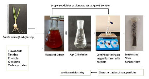
Figure 1: Biological Synthesis of Silver Nanoparticles from Medical Plant Drimia Indica (Roxb.)Jessop
Result and Discussion
UV Visible Spectroscopy
Synthesis of nanomaterials and their applications are having great importance in modern era. Silver nanoparticles were formed by gradual reduction of silver ions during reaction with D.indica plant extract. The reaction mixture showed the color change from colorless to light brown to dark brown.
The initial formation of silver nanoparticles was determined by UV visible spectroscopy. The percentage of transmittance light radiation determines when light of certain frequency is passed through the samples. The spectrophotometer analysis records the intensity of absorption (A) or optical density (O.D) as a function of wavelength. The reaction mixture was taken in quartz cuvette and exposed to UV visible radiation (200 nm-800 nm) to MultiscanSky_1530-00496C spectrophotometer. The synthesized nanoparticles were well dispersed in the solution and were stable for long time by showing brown color to reaction mixture. The absorbance peak wasrecorded for nanoparticles which ranged from 2 nm to 100 nm in size. The absorbance maxima were recorded at 421 nm which clearly indicated the formation of silver nanoparticles (Figure 2).
SEM
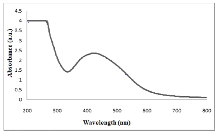
Figure 2: UV-vis absorption spectrum of Silver nanoparticles
The morphology and size of silver nanoparticles derived from D.indica leaf extract were characterized by Scanning Electron Microscope (Figure 3). The image magnification was 10,000 X. The studies revealed the average size of silver nanoparticles was between 50 nm-80 nm. These synthesized nanoparticles were highly stable in nature. The nanoparticles were spherical in shape and agglomerated due to isoelectric pH where, the attraction between the particles reaches a maximum, so it is easy to form hard agglomerates.
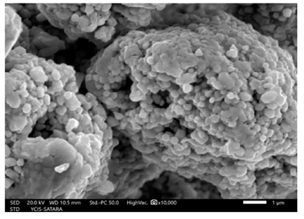
Figure 3: SEM micrograph of Silver nanoparticles.
XRD
To investigate the crystalline nature and structure of Ag NPs, the sample was analyzed on BRUKER-D8 ADVANCE machine at CFC, Shivaji University, Kolhapur. The X-ray diffraction pattern obtained for the silver nanoparticles synthesized from D.indica leaf extract showed five distinct diffraction peaks of 2θ values of 38.11° (111), 44.30°(200), 64.22° (220), 77.51° (311) and 81.47° (222) with the assigned planes indicated in the bracktes. The free crystalline nature of synthesized nanoparticles was done on the basis of JCPDS file. Analysis was done by using Origin Pro 8 software (Figure 4).
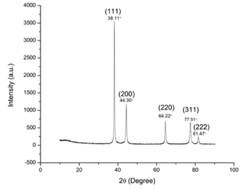
Figure 4: X-ray diffraction pattern of Silver nanoparticles
Antibacterial Assay
The antibacterial activity of silver nanoparticles synthesized from D.indica leaf extract was tested on pathogenic bacteria like P.aeruginosa [ATCC2021], Klebsiella pneumonia [ATCC2075] and E.coli [ATCC2065]. Bacterial cultures were maintained in liquid nutrient broth. Calculation of inhibition activity of synthesized nanoparticles on selected pathogenic bacterial cultures was done using different concentrations of Ag NPs. Antibacterial assay was carried out in 96-well microtiter plate. The assay volume was set to 300 μl which contained 260 μl nutrient broth, 20 μl bacterial culture and 20 μl Ag NPs. The Ag NPs were used in varied concentrations (25 μl/ml, 50 μl/ml and 100 μl/ml from mg/ml stock). Replacement of 20 μl Ag NPs with 20 μl distilled water in assay mixture served as negative control. The experiment was performed in triplicates. The 96-well micro-titre plate was incubated for 12 hours. The readings were recorded after time interval of every 30 minutes in MultiscanSky_1530-00496C spectrophotometer. Graph was plotted which indicated sigmoid growth curve. Similar assay was repeated for all bacteria under study. The lowest concentration which inhibits the growth of bacterial strain is taken as MIC of the Ag NPs for the pathogenic bacteria. Broad spectrum antibiotic streptomycin in different concentrations (25 μg/ml, 50 μg/ml and 100 μg/ml) was used to calculate the MIC values for bacterial strains against streptomycin.
When Ag NPs were tested for antimicrobial activity, P.aeruginosa showed MIC value of 25 μg/ml, K.pneumoniae showed 25 μg/ml and E.coli showed 50 μg/ml (Table 1).
| Sr. No. |
Bacteria |
MIC values µg/ml |
MIC values µg/ml |
| Ag NP’s |
Streptomycin |
| 1 |
Pseudomonas aeruginosa |
25 |
100 |
| 2 |
Klebsiella pneumonia |
25 |
50 |
| 3 |
Escherichia coli |
50 |
100 |
Table 1: MIC values of Silver nanoparticles with Streptomycin.
P.aeruginosa inhibition percentage was recorded to be highest (68%) at 100 μg/ml concentration of streptomycin while it was found to be lowest (45%) at 25 μg/ml Streptomycin concentration. The highest inhibition percentage (65%) at 100 μg/ml and lowest (43%) at 25 μg/ml was recorded for K.pneumoniae. While in case of E.coli antibiotic Streptomycin showed highest inhibition percentage (69%) at 100 μg/ml and lowest inhibition percentage (47%) at 25 μg/ml (Figure 5).
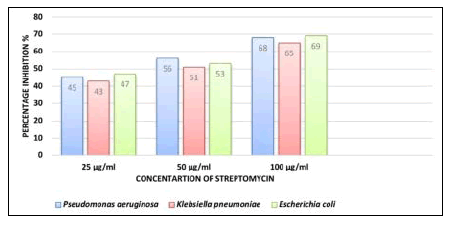
Figure 5: Percentage inhibition of Bacterial species using antibiotic Streptomycin
The highest inhibition percentage of Ag NP’s against P.aeruginosa was observed to be 54% at Ag NPs concentration 100 μg/ml and lowest 39% at 25 μg/ml. In case of K.pneumoniae highest inhibition percentage (49%) was observed at 100 μg/ ml of concentration Ag NP’s and lowest was 35% at 25 μg/ml. While, highest percentage inhibition for E.coli was 51% at 100 μg/ml and lowest was 37% at 25 μg/ml concentration of Ag NPs (Figure 6).
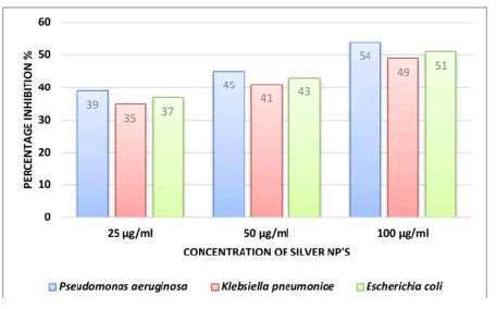
Figure 6: Percentage inhibition of Bacterial species using silver NP’s of D.indica
Thus, while studying the antibacterial activity of P.aeruginosa, highest inhibition percentage (68%) was recorded when Streptomycin (100 μg/ml) was used and when Streptomycin was replaced with same concentration of Ag NPs, the highest inhibition percentage recorded was 54%. In case of K.pneumoniae, highest inhibition percentage (65%) was recorded in Streptomycin (100 μg/ml). The same concentration of Ag NPs exhibited 49% inhibition. Highest inhibition percentage (69%) was recorded in E.coli for Streptomycin (100 μg/ml). Highest inhibition percentage 5% was recorded for E.coli when Ag NP’s (100 μg/ml) was used.
The results depicted that the role of antibiotic was remarkable in inhibition of growth in P.aeruginosa, K.pneumoniae and E.coli. Comparative results avoiding antibiotic and replacing it with biologically synthesized Ag NP’s showed the presence of inhibition activity in all the bacteria tested. Although inhibition activity of Ag NPs was lower than that of antibiotic, the Ag NPs were able to show antibacterial activity. Further research needs to be refined by designing the protocol in which higher concentrations must be tried to determine antibacterial activity. However, high doses of Ag NPs have advantage over higher doses of antibiotics as Ag NPs are eco-friendly, nontoxic and biologically synthesized. Biologically synthesized nanoparticles can be used as better options over hazardous antibiotics by using them as antibacterial agents.
Conclusion
Ag NPs of D.indica has shown the lowest antibacterial effect
than control in present study, but when the concentration
were increased antibacterial effect also increased. It clearly revealed
that the inhibition activity of Ag NPs is completely dose
dependent and it inhibits the pathogenic bacteria. Thus, they
can be widely used as medicines for bacterial diseases. This
will eventually help to investigate the problems and mutations
caused by high doses of antibiotics.
Acknowledgement
Author PK is thankful to BARTI, Pune for financial support and
Head Department of Botany, SUK for providing laboratory facilities.
Conflict of Interest
The author’s declared that they have no conflict of interest.
References
- Thakkar KN, Mhatre SS, Parikh RY (2010) Biological synthesis of metallic nanoparticles. Nanomed Nanotechnol Biol Med. 6(2):257-62.
[Crossref] [PubMed]
- Biological synthesis of metallic nanoparticles
- Pal S, Yu KT, Joon MS (2020) Does the antibacterial activity of silver nanoparticles depend on the shape of the nanoparticle? A study of the gram-negative bacterium escherichia coli. Appl Environ Microbiol. 73(6):1712-20.
[Crossref] [PubMed]
- Emma W, Antoin La, Aine W, Fiona R (2008) The use of nanoparticles in anti-microbial materials and their characterization. Analyst. 133(7):835-45.
[Crossref] [PubMed]
- Linlin WCH, Longquan S (2017) The antimicrobial activity of nanoparticles: Present situation and prospects for the future. Int J Nanomedicine. 12:1227-1249.
[Crossref] [PubMed]
- Heather K. Allen (2017) Alternatives to antibiotics: Why and how. Natl Acad Med.
[Crossref] [Google Scholar]
- Jayanta KP, Gitishree D, Leonardo FF, Estefania VRC, Maria DPRT, et al. (2018) Nano based drug delivery systems: Recent developments and future prospects. J Nanobiotechnol. 16(1):71.
[Crossref] [PubMed]
- Marx J, Pretorius E, Espag WJ, Bester MJ (2005) Urginea sanguinea: Medicinal wonder or death in disguise? Environ Toxicol Pharmacol. 20(1):26-34.
[Crossref] [PubMed]
- Crespo MB, Martínez AM, Alonso MÁ (2020) The identity of Drimia purpurascens, with a new nomenclatural and taxonomic approach to the “Drimia undata” group (Hyacinthaceae=Asparagaceae subfam. Scilloideae). Plant Syst Evol. 306(67):1–18.
[Crossref] [Google Scholar]
- Chittoor MS, Binny AJR, Yadlapalli SK, Cheruku A, Dandu C, et al. (2012) Anthelmintic and antimicrobial studies of Drimia indica (Roxb.) Jessop. bulb aqueous extracts. J Pharm Res. 5(5):3677-3686.
[Google Scholar]
- Pandey D, Gupta AK (2014) Antimicrobial activity and phytochemical analysis of Urginea indica from Bastar district of Chhattisgarh. Int J Pharm Sci Rev Res. 26(2):273-281.
[Google Scholar]
- Sonali A, Ankit K, Ruchi BS, Ashutosh C, Abhimanyu K, et al. (2019) Drimia indica: A plant used in traditional medicine and its potential for clinical uses. Medicina (Kaunas). 55(6):255.
[Crossref] [PubMed]
- Madira CM, Gothusaone ST, Given TM, Keamogetswe PS, John FM, et al. (2021) Bulbous plants Drimia: “A thin line between poisonous and healing compounds” with biological activities. Pharm. 13(9):1385.
[Crossref] [PubMed]
Citation: Kamble PS, Gadade JP, Patil DS, Jagtap MA, Jayannawar DP, et al. (2023) Biosynthesis of Silver Nanoparticles Using Drimia indica and Exploring its Antibacterial Profile. J Nanosci Nanotechnol Res. 7.01.
Copyright: © 2023 Kamble PS, et al. This is an open-access article distributed under the terms of the Creative Commons Attribution
License, which permits unrestricted use, distribution, and reproduction in any medium, provided the original author and
source are credited.


