Keywords
Carcinoma, Pancreatic Ductal; Micro RNAs; MIRN21 Micro RNA, human; MIRN155 Micro RNA, human; Pancreatic Juice
INTRODUCTION
Pancreatic ductal adenocarcinoma is the fifth leading cause of cancer deaths in Japan and fourth in the United States. Chemoradiotherapy has been shown to prolong survival time in recent years; however, the prognosis remains extremely poor with a median survival time of less than 24 months [1, 2]. The poor prognosis of pancreatic ductal adenocarcinoma is due to the difficulty of achieving an early diagnosis in addition to its aggressive malignant behavior. Endoscopic retrograde cholangiopancreatography (ERCP) and subsequent cytological assessment of pancreatic juice is frequently used to diagnose pancreatic ductal adenocarcinoma; however, the sensitivity of cytological assessment has been reported to range from 40 to 60% [3, 4]. Therefore, the identification of unique molecules in pancreatic juice targeting pancreatic ductal adenocarcinoma is necessary for an early and definitive diagnosis of pancreatic ductal adenocarcinoma.
Micro RNAs (miRs) are 18-24 nucleotide RNA molecules which modulate the translation of specific mRNAs and play an important role in a wide range of physiologic and pathologic processes [5]. Several recent studies have identified the overexpression of miRs in pancreatic ductal adenocarcinoma by the use of resected specimens. Szafranska et al. [6] have studied the expression profiling of miRs using resected specimens of pancreatic ductal adenocarcinoma and have demonstrated that a total of eleven miRs including miR-216 and miR-217 were downregulated while six miRs, such as miRs-205, -18a, -31, -93, -221, and -224, were overexpressed in pancreatic ductal adenocarcinoma. Wang et al. [7] analyzed the expression of miRs in the plasma of patients with pancreatic ductal adenocarcinoma and identified miRs- 21, -210, -155, and -196a as candidate biomarkers for the detection of pancreatic ductal adenocarcinoma. Bloomston et al. [8] showed that miR-21 and miR-155 were overexpressed in pancreatic cancer as compared to chronic pancreatitis, and we also found high expression levels of miR-21 in pancreatic cancer cells [9]. Habbe et al. [10] reported that miR-155 was postoperatively. overexpressed in the cystic contents of intraductal papillary mucinous neoplasms of the pancreas (IPMN) On the other hand, the expression of miRs in pancreatic juice has not been assessed to date in patients with pancreatic ductal adenocarcinoma.
In this preliminary study, we focused on miR-21 and miR-155, based on the previous findings as described above, and evaluated the expression levels of these miRs in pancreatic juice in patients with pancreatic ductal adenocarcinoma.
MATERIALS AND METHODS
Formalin-fixed paraffin-embedded tissue samples and pancreatic juice samples were collected from patients treated at our hospital between 2006 and 2009. The diagnosis of pancreatic ductal adenocarcinoma was determined by the pathological results of resected specimens while that of chronic pancreatitis was made based on the pathology of the resected specimens or clinical findings at the time of initial diagnosis and during a follow-up period of at least 12 months.
Tissue Samples
Formalin-fixed paraffin-embedded tissue samples were collected from 9 patients with pancreatic ductal adenocarcinoma and three patients with chronic pancreatitis who underwent pancreatectomy. One normal pancreatic sample was also collected from the normal pancreas tissue of one patient with PDAC; therefore, 4 control samples were available. Using section slides having a 10 μm thickness, macrodissection was performed as previously reported [11].
Pancreatic Juice Samples
Pancreatic juice samples were obtained from 16 patients with pancreatic ductal adenocarcinoma and 5 with chronic pancreatitis. A 5-Fr wedge pressure balloon catheter was inserted into the main pancreatic duct during ERCP, and the duodenoscope was then removed from the patient, leaving the catheter in place. Pancreatic juice was collected through the catheter for 10 to 20 minutes after an intravenous administration of 1 μg of secretin (ChiRhoClin, Inc., Burtonsville, MD, USA) dissolved in 5 mL of 0.9% saline. About 10 to 20 mL of pancreatic juice were usually collected by this method, and half of the pancreatic juice collected was sent to the pathological unit for cytology. The other half was centrifuged for 20 minutes at 3,000 rpm at 4°C, and the pellet was separated from the supernatant and stored at -20°C for later molecular analyses.
Isolation of RNA
Total RNA was extracted from the cell pellets of pancreatic juice according to the standard guanidinium thiocyanate-phenol-chloroform protocol with glycogen (mirVana PARIS Kit.; Applied Biosystems, Carlsbad, CA, USA), and from sections isolated by macrodissection with a PicoPure RNA Isolation Kit (Arcturus Bioscience, Mountain View, CA, USA) with DNase I treatment according to the manufacturer’s instructions [11]. The concentration of RNA was measured with a NanoDrop ND-1000 spectrophotometer (NanoDrop Technologies, Wilmington, DE, USA), determining absorbance at 260 and 280 nm. RNA integrity was assessed with an Agilent Bioanalyzer 2100 (Agilent, Palo Alto, CA, USA).
Quantitative Real-Time Reverse Transcription-PCR Assay for miR-21, miR-155, and miR-191
Quantitative real-time reverse transcription-PCR (qRTPCR) was performed with a Chromo4 Real-Time PCR Detection System (Bio-Rad Laboratories, Hercules, CA, USA) using a TaqMan® Micro RNA Reverse Transcription Kit and TaqMan® Universal PCR Master Mix (Applied Biosystems, Carlsbad, CA, USA), in accordance with the manufacturer’s instruction [9]. For the measurement of miR-21, miR-155, miR-191 and RNU6B expression (U6 snRNA, a reference gene), we performed two-step qRT-PCR with specific primers for miR-21, miR-155 and RNU6B (designed by Applied Biosystems, Carlsbad, CA, USA), following the manufacturer’s protocol. Briefly, the reaction mixture was initially incubated at 16°C for 30 min, 42°C for 30 min and 85°C for 5 min, and then held at 4°C for the reverse transcription step until the preparation for the PCR reaction had been completed. PCR amplification was initiated with one cycle of 95°C for 10 min to activate the AmpliTaq Gold® Enzyme (Applied Biosystems, Carlsbad, CA, USA), followed by 40 cycles of 94°C for 15 s and 60°C for 60 s. Each sample was run in triplicate wells. The levels of miR-21 and miR-155 expression were calculated from a standard curve constructed with small RNAs from the Suit-2 pancreatic cancer cell line. The miR-21 and miR-155 expression levels were stable and constant in the Suit-2 pancreatic cell line. We used the dCT method to calculate the miR expression. Normalizations were carried out with small nuclear RNA U6 (RNU6B; Applied Biosystems, Carlsbad, CA, USA) or miR-191 [12]. Peltier and Latham [12] reported that miR-191 is one of the most stable micro RNAs. Their data suggest that miR-191 is more likely to be a reasonable choice than the default use of U6 and 5S.
ETHICS
Written informed consent was obtained from all patients, the study was approved by ethical and surveillance committees of Kyushu University Hospital and conducted according to the Helsinki Declaration.
STATISTICS
Statistical analysis was performed with the JMP 7.0.1 (SAS Institute, Inc., Cary, NC, USA). The Mann- Whitney U test was utilized to evaluate the differences between the two groups. Differences were considered significant when the two-tailed P value was less than 0.05.
RESULTS
Expression of RNA U6 and miR-191 in Pancreatic Juice Samples
Sixteen pancreatic juice samples obtained from patients with pathologically confirmed pancreatic ductal adenocarcinoma were analyzed for the expression of RNA U6 and miR-191 to assess the quality of our experiments. As shown in Figure 1, miR-191 was detectable in all the samples. On the other hand, RNA U6 was detected in 12 of the 16 samples. These 12 samples were used for the additional analyses of miR- 21 and miR-155 expression in pancreatic ductal adenocarcinoma patients (sample number 1-12). Three of the 5 pancreatic juice samples from chronic pancreatitis patients were available for the additional analyses while the other two were not because miR- 191 was not detected.
Figure 1. Expressions of RNA U6 (U6) and microRNA-191 (miR-
191) in pancreatic juice samples. The samples were obtained from 16
patients with pathologically confirmed pancreatic ductal adenocarcinoma
(PDAC) and from 5 patients with chronic pancreatitis
(CP). MicroRNA-191 was detectable in all pancreatic ductal
adenocarcinoma samples and 3 chronic pancreatitis samples while
RNA U6 was detected in 12 of the 16 pancreatic ductal
adenocarcinoma samples and 3 of the 5 chronic pancreatitis samples.
Sample numbers (1-15) correspond to the sample numbers in Table 1.
N/D: not detectable.
Ct: threshold cycle
Expression of miR-21 and miR-155 in Formalin- Fixed Paraffin-Embedded and Pancreatic Juice Samples
Figure 2 demonstrates that the relative expression levels of miR-21 in formalin-fixed paraffin-embedded (P=0.009) and pancreatic juice samples (P=0.021) were significantly higher in patients with pancreatic ductal adenocarcinoma as compared to those in the controls (chronic pancreatitis and normal pancreas).
Figure 2. Expression of microRNA-21 in formalin-fixed paraffinembedded
(FFPE) tissue and pancreatic juice samples is shown in a
box-and-whisker diagram, which presents the smallest and largest
observations (except outlier(s); box) and lower quartile, median,
upper quartile (whiskers). Relative expression levels of microRNA-
21 in tissue (P=0.009) and pancreatic juice samples (P=0.021) were
significantly higher in patients with pancreatic ductal
adenocarcinoma (PDAC) as compared to the controls: chronic
pancreatitis (CP) and normal pancreas (NP).
Figure 3 shows that the relative expression levels of miR-155 in formalin-fixed paraffin-embedded (P=0.014) and pancreatic juice samples (P=0.021) were significantly higher in patients with pancreatic ductal adenocarcinoma as compared to those in the controls (chronic pancreatitis and normal pancreas).
Figure 3. Expression of microRNA-155 in formalin-fixed paraffinembedded
(FFPE) tissue and pancreatic juice samples is shown in a
box-and-whisker diagram, which presents the smallest and largest
observations (except outlier(s); box) and lower quartile, median, and
upper quartile (whiskers). Relative expression levels of microRNA-
155 in tissue (P=0.014) and pancreatic juice samples (P=0.021) were
significantly higher in patients with pancreatic ductal
adenocarcinoma (PDAC) as compared to the controls: chronic
pancreatitis (CP) and normal pancreas (NP).
Relationship between Pancreatic Juice Cytology and Expression Levels of miRs
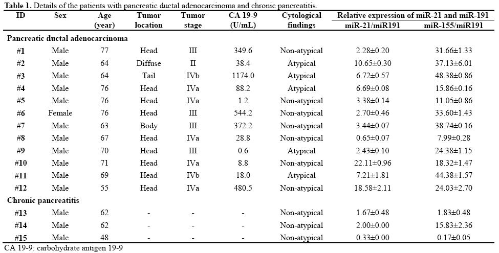
The presence of atypical cells (class III-IV-V) in pancreatic juice samples was found in 5 of 12 patients with pancreatic ductal adenocarcinoma (41.7%, 3 of class III, 2 of class IV, and none of class V) (Figure 4). The rtelationship between the pancreatic juice cytology and the expression levels of miRs in 12 patients having pancreatic ductal adenocarcinoma are shown in Figure 5. Expression levels of miR-21 in pancreatic juice of patients with a cytological result of non-atypical cells (no. 7, 58.3%; all of class II) were not significantly different from those of the atypical results (P=0.246). The same finding was observed in the expression levels of miR-155 in pancreatic juice between non-atypical and atypical results (P=0.592). Table 1 shows the details of the samples.
Figure 4. a. Non-atypical cells in pancreatic juice (x400). b. Atypical
cells in pancreatic juice (x400).
Figure 5. the relationship between pancreatic juice cytology and
expression levels of microRNAs in 12 patients with pancreatic ductal
adenocarcinoma is shown in a box-and-whisker diagram, which
presents the smallest and largest observations (except outlier(s); box)
and lower quartile, median, and upper quartile (whiskers). The
expression level of microRNA-21 (miR-21) in patients with nonatypical
results (no. 7) was not different from that of the atypical
results (no. 5). The same result was obtained in the expression levels
of microRNA-155 (miR-155) in pancreatic juice between nonatypical
and atypical cytology results.
DISCUSSION
Our present study analyzing the expression levels of miRs in pancreatic ductal adenocarcinoma tissue samples and pancreatic juice collected by preoperative ERCP demonstrated several informative results [1]. MiRs were detectable in pancreatic juice [2]. The expression levels of miR-21 and miR-155 in pancreatic ductal adenocarcinoma were significantly higher than those in chronic pancreatitis, and the same results were obtained in formalin-fixed paraffin-embedded samples. [3] The expression levels of miR-21 and miR-155 were not correlated with the results of pancreatic juice cytology.
Direct pancreatography for the morphological evaluation of the pancreatic duct and subsequent pancreatic juice collection for cytology are the gold standard for the diagnosis of pancreatic ductal adenocarcinoma; however, the sensitivity of cytological assessment has been unsatisfactory [3, 4]. Therefore, several analyses of pancreatic juice samples to detect the new molecules have been attempted to date in order to increase the possibility of diagnosing pancreatic ductal adenocarcinoma [11, 13]. MiRs have recently gained attention as being new biomarkers for various malignant diseases, including pancreatic ductal adenocarcinoma [6, 7, 8, 9, 14, 15]. Previous reports have demonstrated that specific miRs in resected specimens or in serum samples obtained postoperatively from patients with pancreatic ductal adenocarcinoma and in cystic contents of IPMNs [6, 7, 8, 9, 10] could become new markers for the diagnosis of pancreatic ductal adenocarcinoma and IPMNs. On the other hand, as miRs in pancreatic juice in patients with pancreatic ductal adenocarcinoma have not been assessed to date, this is the first report which evaluates the expression of miRs in pancreatic juice preoperatively and demonstrates the high expression levels of some specific miRs in pancreatic juice of pancreatic ductal adenocarcinoma patients as compared to those of chronic pancreatitis patients. MiR-21 is one of the most frequently examined for various malignant diseases including pancreatic ductal adenocarcinoma and cancers of the lung, stomach, breast, colon, and prostate [7, 8, 9, 14, 15, 16, 17]. We have recently shown that miR-21 enhances proliferation, invasion and gemcitabine-resistance of pancreatic cancer cells by affecting matrix metalloproteinase-2 and -9 and vascular endothelial growth factor expressions [9]. Chan et al. [16] demonstrated that miR-21 plays an important role in preventing apoptosis using glioblastoma cell lines. The present study also showed higher expression levels of miR-21 in tissue samples of pancreatic ductal adenocarcinoma as compared to those of chronic pancreatitis and, therefore, the expression levels of miR-21 in pancreatic juice were expected to reflect the miR-21 levels in main tumors.
MiR-155 has been reported to be associated with activation of lymphoma cells, such as diffuse large cell, Hodgkin’s and Burkitt types [18, 19]. Others have shown that miR-155 regulates T-cell activation and immune responses [20, 21]. Therefore, the main function of miR-155 might be the regulation of lymphocytes. Although the role of miR-155 in carcinogenesis or cancer progression has not been adequately understood, several articles have reported high expression levels of miR-155 in pancreatic ductal adenocarcinoma [7, 8]. Additional studies are necessary to clarify the significance of miR-155 expression in pancreatic ductal adenocarcinoma and its diagnostic application using pancreatic juice.
Atypical cells in pancreatic juice were detected in only 42% of the patients with pancreatic ductal adenocarcinoma in the present study, similar to previous studies reporting values from 40 to 60% [3, 4]. The expression levels of miR-21 and miR-155 in pancreatic juice were not different between pancreatic ductal adenocarcinomas presenting positive and negative preoperative cytological results but could significantly distinguish pancreatic ductal adenocarcinoma from chronic pancreatitis. The assessment of the expression levels of miRs in pancreatic juice would have a potential as markers for the diagnosis of pancreatic ductal adenocarcinoma even in the absence of atypical cells in pancreatic juice. The sample number might not be sufficient to argue this relationship. Additional studies are required to determine the cut-off value of the expression levels of these miRs in pancreatic juice and to clarify the diagnostic value of the combination of the expression levels of these miRs with cytological results to increase the diagnostic potential of miRs.
Conflict of interest The authors have no potential conflict of interest
References
- Hirata K, Egawa S, Kimura Y, Nobuoka T, Oshima H, Katsuramaki T, et al. Current status of surgery for pancreatic cancer. Dig Surg 2007; 24:137-47. [PMID 17476103]
- Jemal A, Siegel R, Ward E, Hao Y, Xu J, Thun MJ. Cancer statistics, 2009. CA Cancer J Clin 2009; 59:225-49. [PMID 19474385]
- Fujita N, Noda Y, Kobayashi G, Kimura K, Ito K. Endoscopic approach to early diagnosis of pancreatic cancer. Pancreas 2004; 28:279-81. [PMID 15084971]
- Wakatsuki T, Irisawa A, Bhutani MS, Hikichi T, Shibukawa G, Takagi T, et al. Comparative study of diagnostic value of cytologic sampling by endoscopic ultrasonography-guided fine-needle aspiration and that by endoscopic retrograde pancreatography for the management of pancreatic mass without biliary stricture. J Gastroenterol Hepatol 2005; 20:1707-11. [PMID 16246190]
- Pillai RS. Micro RNA function: multiple mechanisms for a tiny RNA? RNA 2005; 11:1753-61. [PMID 16314451]
- Szafranska AE, Davison TS, John J, Cannon T, Sipos B, Maghnouj A, et al. Micro RNA expression alterations are linked to tumorigenesis and non-neoplastic processes in pancreatic ductal adenocarcinoma. Oncogene 2007; 26:4442-52. [PMID 17237814] 7
- . Wang J, Chen J, Chang P, LeBlanc A, Li D, Abbruzzesse JL, et al. Micro RNAs in plasma of pancreatic ductal adenocarcinoma patients as novel blood-based biomarkers of disease. Cancer Prev Res (Phila) 2009; 2:807-13. [PMID 19723895]
- Bloomston M, Frankel WL, Petrocca F, Volinia S, Alder H, Hagan JP, et al. Micro RNA expression patterns to differentiate pancreatic adenocarcinoma from normal pancreas and chronic pancreatitis. JAMA 2007; 297:1901-8. [PMID 17473300]
- Moriyama T, Ohuchida K, Mizumoto K, Yu J, Sato N, Nabae T, et al. Micro RNA-21 modulates biological functions of pancreatic cancer cells including their proliferation, invasion, and chemoresistance. Mol Cancer Ther 2009; 8:1067-74. [PMID 19435867]
- Habbe N, Koorstra JB, Mendell JT, Offerhaus GJ, Ryu JK, Feldmann G, et al. Micro RNA miR-155 is a biomarker of early pancreatic neoplasia. Cancer Biol Ther 2009; 8:340-6. [PMID 19106647]
- Ohuchida K, Mizumoto K, Yu J, Yamaguchi H, Konomi H, Nagai E, et al. S100A6 is increased in a stepwise manner during pancreatic carcinogenesis: clinical value of expression analysis in 98 pancreatic juice samples. Cancer Epidemiol Biomarkers Prev 2007; 16:649-54. [PMID 17416753]
- Peltier HJ, Latham GJ. Normalization of Micro RNA expression levels in quantitative RT-PCR assays: identification of suitable reference RNA targets in normal and cancerous human solid tissues. RNA 2008; 14:844-52. [PMID 18375788]
- Nakashima A, Murakami Y, Uemura K, Hayashidani Y, Sudo T, Hashimoto Y, et al. Usefulness of human telomerase reversetranscriptase in pancreatic juice as a biomarker of pancreatic malignancy. Pancreas 2009; 38:527-33. [PMID 19342980]
- Volinia S, Calin GA, Liu CG, Ambs S, Cimmino A, Petrocca F, et al. A Micro RNA expression signature of human solid tumors defines cancer gene targets. Proc Natl Acad Sci USA 2006; 103:2257-61. [PMID 16461460]
- Iorio MV, Ferracin M, Liu CG, Veronese A, Spizzo R, Sabbioni S, et al. Micro RNA gene expression deregulation in human breast cancer. Cancer Res 2005; 65:7065-70. [PMID 16103053]
- Chan JA, Krichevsky AM, Kosik KS. Micro RNA-21 is an antiapoptotic factor in human glioblastoma cells. Cancer Res 2005; 65:6029-33. [PMID 16024602]
- Meng F, Henson R, Lang M, Wehbe H, Maheshwari S, Mendell JT, et al. Involvement of human micro-RNA in growth and response to chemotherapy in human cholangiocarcinoma cell lines. Gastroenterology 2006; 130:2113-29. [PMID 16762633]
- Kluiver J, Poppema S, de Jong D, Blokzijl T, Harms G, Jacobs S, et al. BIC and miR-155 are highly expressed in Hodgkin, primary mediastinal and diffuse large B cell lymphomas. J Pathol 2005; 207:243-9. [PMID 16041695]
- Kluiver J, Haralambieva E, de Jong D, Blokzijl T, Jacobs S, Kroesen BJ, et al. Lack of BIC and Micro RNA miR-155 expression in primary cases of Burkitt lymphoma. Genes Chromosomes Cancer 2006; 45:147-53. [PMID 16235244]
- Lu LF, Thai TH, Calado DP, Chaudhry A, Kubo M, Tanaka K, et al. Foxp3-dependent Micro RNA155 confers competitive fitness to regulatory T cells by targeting SOCS1 protein Immunity 2009; 30:80-91. [PMID 19144316]
- Chen DF, Gong BD, Xie Q, Ben QW, Liu J, Yuan YZ. Micro RNA155 is induced in activated CD4(+) T cells of TNBS induced colitis in mice. World J Gastroenterol 2010; 16:854-61. [PMID 20143464]


