Keywords
Insulinoma; Neuroendocrine Tumors; Pancreatic Neoplasms; Proto- Oncogenes
Abbreviations
AMV: avian myeloblastosis virus; CaSm: cancer associated Sm-like; MEN-1: multiple endocrine neoplasia type 1; NF: non-functioning; PET: pancreatic endocrine tumor; RPMI: Roswell Park Memorial Institute; VHL: von Hippel-Lindau.
INTRODUCTION
Little is known about the molecular alterations leading to pancreatic endocrine tumor (PET) tumorigenesis. However, the emergence of novel molecular characterization strategies has made it apparent that PETs exhibit diverse molecular fingerprints with respect to tumors arising from the exocrine component of the pancreas [1, 2, 3, 4]. The p53, K-ras, p16 and DPC4 genes are only rarely altered in PETs [2, 5] and, to date, alteration of the MEN-1 gene remains the most common genetic event noted in gastrinomas and non-functioning PETs [1, 6, 7, 8, 9, 10, 11]. More recently, it has also been shown that non-functioning (NF) PETs may be divided into two molecular phenotypes, distinguishable by ploidy [4]. When utilized in conjunction with the Ki-67 cellular proliferation index, ploidy analysis provides independent statistically significant information which predicts survival [4].
The cancer associated Sm-like (CaSm) gene codes for a protein containing 133 amino acids which contain two Sm motifs found in common small nuclear ribonucleoproteins and has the greatest homology to the SmG protein [12, 13, 14]. CaSm has recently been found to be overexpressed in 87.5% of pancreatic adenocarcinomas when compared to their normal pancreatic tissue and a variety of different tumor types [13]. In addition, the pancreatic adenocarcinoma cell lines, ASPC- 1, CAPAN-2, COLO357, PANC-1, CAPAN- 1 and HPAC were all found by Northern blot analysis to overexpress CaSm when compared to normal pancreatic tissue [13]. Although the exact mechanism of CaSm proto-oncogene overexpression in oncogenesis is unclear, it is thought that, because of its significant homology to the Sm G protein, it may play a role in RNA splicing [15]. Additional evidence reveals homology of CaSm to the yeast homolog Lsm1 indicating a possible role in mRNA decapping [13, 16]. The importance of CaSm expression in contributing to the transformed state has been manifested by its apparent requirement for transformed pancreatic cell lines to be able to grow in an anchorage-independent manner [13]. In addition, antisense CaSm RNA has been shown to have significant anti-tumor effects at both the cellular and the animal levels [13, 15]. In particular, pancreatic cell lines infected with Ad-alpha-CaSm revealed a statistically significant reduction in proliferation and anchorage independent growth [15]. Using immune-compromized mice with subcutaneous tumor implants, a decrease in tumor growth and increase of median survival was further demonstrated [15]. Because of its potential role in pancreatic oncogenesis, an analysis of Smlike gene expression levels in PETs is also warranted.
Using real-time PCR, we have examined the expression levels of CaSm in a panel of 15 primary and 7 metastatic PETs, and compared expression levels to those seen in normal islet cells.
MATERIALS AND METHODS
Patients and Tumors
Tumors of 17 patients (8 males and 9 females) were studied. The average age of the patients was 52 years (range 28-75). A total of 15 primary pancreatic tumors (including 10 NF-PET and 5 primary insulinomas) and 7 NF-PET hepatic metastases were studied. Some of these cases have been previously described [4, 17]. Five of the patients with metastatic NF cancers also had the corresponding primary tumor available. One NF tumor and one insulinoma were from patients with MEN-1 syndrome. All tumor samples were collected in the operating room in sterile containers and snap frozen in liquid nitrogen. Histology was confirmed on frozen sections and only tumors of more than 90% tumor cells were used. Normal pancreas and islet cells were from organ donors.
Cell Lines
The pancreatic adenocarcinoma cell lines GER, T3M4, CFPAC1, and PANC-2 were obtained from NR Lemoine (Imperial College School of Medicine. London, UK), PACA3, PACA44 from M von Bülow (University of Mainz. Mainz, Germany), PANC-1, ASPC-1 (American Type Culture Collection. Manassas, VA, USA) and A8184 from H Kalthoff (University Hospital Eppendorf. Hamburg, Germany). All the cell lines were cultured in Roswell Park Memorial Institute (RPMI) 1640 medium supplemented with 10% heat-inactivated fetal calf serum (Gibco- BRL, Life Technologies Inc. Frederick, MA, USA) and were Mycoplasma free.
RNA Extraction and cDNA Synthesis
Total RNA from tissue specimens and cell lines was prepared using TRIZOL reagent (Gibco-BRL, Life Technologies Inc. Frederick, MA, USA) according to the manufacturer’s protocol. Complementary DNA was synthesized with the 1st Strand cDNA Synthesis Kit for RT-PCR using avian myeloblastosis virus (AMV) reverse transcriptase and oligo (dT) primers using the manufacturer’s recommendations (Roche Molecular Biochemicals. Mannheim, Germany).
Real-Time PCR
The sense and antisense primers used were TACATATGCAACATGAAGAA and CTCTAATTTGGGATATGAAG, respectively, which recognize sequences near the 5' end of the cDNA and amplify a 150-bp fragment. The PCR conditions were 95 °C for 10 min, followed by 40 cycles of 95 °C for 45 sec, 58 °C for 45 sec, and 72 °C for 45 sec. PCR reactions contained 5 μL of SYBER Green PCR Master Mix (PE Biosystems. Foster City, CA, USA), 10 ng of primers and 500 ng of cDNA in a total volume of 10 μL and were performed in triplicate. Beta-actin was also amplified under the same conditions and used to normalize reactions. Sense and antisense primers to the beta-actin gene were TGGAGAAAATCTGGCACCACACC and GATGGGCACAGTGTGGGTGACCC, respectively.
A 9600 thermocycler mounted with an ABI Prism 7700 sequence detection system (Perkin Elmer Biosystems. Foster City, CA, USA), was used to perform the PCR cycles and fluorescence was quantified as a Ct value with Primer Express software (Perkin Elmer Biosystems. Foster City, CA, USA). CaSm expression levels were calibrated using betaactin expression levels as an internal control. The differences between the mean Ct values of the gene of interest and the housekeeping gene were denoted (delta-Ct) and the difference between delta-Ct and the Ct value of the calibrator sample was labeled deltadelta- Ct. The log2(delta-delta-Ct) gave the relative quantitation value of CaSm expression with islet cell expression designated as one. Control wells containing SYBER Green PCR master mix and primers without sample cDNA emitted no fluorescence after 40 cycles.
ETHICS
All the studies performed were approved by the Ethics Committee of Verona University, and the study protocol conforms to the ethical guidelines of the 1975 Declaration of Helsinki.
STATISTICS
Descriptive statistics were evaluated and the frequencies of cases which showed elevated expression levels of CaSm (defined as a twofold, or greater value when compared to expression levels found in normal pancreas obtained from healthy donors) were reported.
RESULTS
Since it has previously been shown that pancreatic cancer cell lines overexpress CaSm when compared to normal pancreatic tissue [13], a panel of nine pancreatic adenocarcinoma cell-lines was used as positive controls for the ability to detect and quantitate CaSm mRNA levels using realtime PCR. Beta-actin was used to normalize expression levels. The results of this analysis are detailed in Table 1. Five of these cell-lines (56%) showed elevated expression levels (two-fold or greater) when compared to expression levels found in normal pancreas obtained from healthy donors. Thus, these results confirm a previous report which demonstrated elevated expression of CaSm in pancreatic cancer cell-lines [13] and further demonstrate the validity of the experimental system.

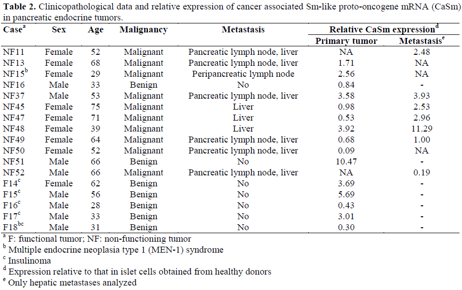
We next sought to quantify the levels of expression of CaSm in pancreatic endocrine tumors. Pertinent clinicopathological characteristics of the cases under study are detailed in Table 2. A total of 10 NF-PETs, 5 insulinomas and 7 NF hepatic metastases were analyzed. The expression levels of CaSm were normalized to those found in islet cells isolated from a normal pancreas of an organ donor. Four of the 10 NF tumors (40%) and 3 of the 5 (60%) insulinomas showed elevated expression levels of CaSm. The gene was also overexpressed in 5 of the 7 metastatic PETs analyzed (71%).
DISCUSSION
The yearly incidence of pancreatic cancer in the United States is around 30,000 and it is the fifth leading cause of cancer-related death; in Europe, it causes 30,000 deaths per year. The vast majority of pancreatic cancers are ductal in origin with non-ductal pancreatic cancer comprising about 10% of all tumors diagnosed. Of these, about half are pancreatic endocrine tumors which arise from pancreatic islet cells and may occur either sporadically or as part of multiple endocrine neoplasia type 1 (MEN-1) or von Hippel-Lindau (VHL) syndromes. PETs are traditionally divided into functional and non-functional subtypes depending on whether or not the tumors secrete hormones that give rise to clinical symptoms. Functional PETs are mainly represented by insulinomas, are usually smaller and have a more favorable clinical outcome with respect to NF-PETs; it is possible that this is a consequence of the hormone related symptoms which allow a more timely diagnosis of functional PETs. In fact, the initial clinical presentation of NFPET is commonly an abdominal mass or any symptom related to the invasion of adjacent structures. PETs are generally slow growing with an average delay in diagnosis of five years and have a higher degree of resectability in contrast to their exocrine counterparts. Because symptoms can usually be controlled pharmacologically (i.e., octreotide, omeprazole) when tumors are functional, the most common cause of death is from liver failure secondary to hepatic metastasis [18]. As CaSm has been reported to be overexpressed in a wide variety of cancers originating from different tissues including that of the pancreas, liver, ovary, lung and kidney [13], it was of interest to determine if it is also overexpressed in pancreatic tumors other than those arising from the pancreatic ducts. In fact, when grouped together, roughly one-half of all primary PETs revealed overexpression of CaSm. Sixty percent of insulinomas also had CaSm overexpression. Most insulinomas are benign with malignancy rates ranging from 4 to 16% [19] and, in our small panel of five tumors, none were found to have evidence of metastasis or local invasion. Unfortunately, patient NF15 also happened to be one of only two patients with the MEN-1 syndrome making any comments as to the relevance of CaSm expression and this syndrome even more difficult.
Of particular interest, overexpression of the gene was found in 5 of the 7 metastatic PETs. When the tumors from the 5 patients with both primary and metastatic samples were compared, it was noteworthy that all of the secondary tumors expressed more of the proto-oncogene than their primary counterparts (Figure 1). It would be tempting to speculate that overexpression of this gene is more frequent in NF metastatic PETs; however, the number of cases analyzed is too small to make any definitive conclusions. This was exacerbated by the fact that material from 2 patients with hepatic metastases (NF13 and NF50) and one patient with a lymph node metastasis (NF15) was not available for examination. Our results, therefore, seem to indicate that CaSm overexpression may have more of a role in PET tumorigenesis than carcinogenesis. This is reiterated by the observation that patient NF51 has one of the highest levels of CaSm expression yet has no evidence of metastatic disease.
Figure 1. Relative cancer-associated Sm-like protooncogene
(CaSm) expression from primary and
secondary tumors of five patients with non-functioning
pancreatic endocrine tumors compared to expression
levels from islets of Langerhans from normal donors.
Nonetheless, as CaSm expression appears to be necessary for the maintenance of the transformed state, both in cellular and animal models of pancreatic adenocarcinoma, it has recently been demonstrated that gene therapy approaches using antisense CaSm RNA have antitumor effects [15]. As a result, this gene may represent a novel gene target for new treatment approaches in primary and metastatic PETs that overexpress this protooncogene. Studies with a larger patient population are needed to better assess this; however, the rarity of these tumors currently limits our ability to do so. Future studies in CaSm protein production in PETs and other pancreatic tumors may help further clarify the role of this gene in pancreatic oncogenesis.
Acknowledgement
Supported by: Fondazione cassa di Risparmio di Verona (bando 2001), Associazione Italiana Ricerca Cancro (AIRC) to A Scarpa, Milan, Italy; Consorzio Studi Universitari di Verona, Verona, Italy; Ministero Italiano Università e Ricerca (Cofin MM06158571, 9906218982), Rome, Italy; Ministero Sanitá (Ricerca finalizzata ICS060.2/RF00-57), Rome, Italy. AA Gumbs was supported by a research fellowship from Ministero Italiano Università e Ricerca, Rome, Italy.
References
- Moore PS, Missiaglia E, Antonello D, Zamo A, Zamboni G, Corleto V, et al. Role of disease-causing genes in sporadic pancreatic endocrine tumors: MEN1 and VHL. Genes Chromosomes Cancer 2001; 32:177- 81. [AN 21433812]
- Moore PS, Orlandini S, Zamboni G, Capelli P, Rigaud G, Falconi M, et al. Pancreatic tumours: molecular pathways implicated in ductal cancer are involved in ampullary but not in exocrine nonductal or endocrine tumorigenesis. Br J Cancer. 2001; 84:253- 62. [AN 21094882]
- Moore PS, Zamboni G, Brighenti A, Lissandrini D, Antonello D, Capelli P, et al. Molecular characterization of pancreatic serous microcystic adenomas: evidence for a tumor suppressor gene on chromosome 10q. Am J Pathol 2001; 158:317-21. [AN 20580919]
- Rigaud G, Missiaglia E, Moore PS, Zamboni G, Falconi M, Talamini G, et al. High resolution allelotype of nonfunctional pancreatic endocrine tumors: identification of two molecular subgroups with clinical implications. Cancer Res 2001; 61:285-92. [AN 21036722]
- Pellegata NS, Sessa F, Renault B, Bonato M, Leone BE, Solcia E, Ranzani GN. K-ras and p53 gene mutations in pancreatic cancer: ductal and nonductal tumors progress through different genetic lesions. Cancer Res 1994; 54:1556-60. [AN 94184977]
- Cupisti K, Hoppner W, Dotzenrath C, Simon D, Berndt I, Roher HD, Goretzki PE. Lack of MEN1 gene mutations in 27 sporadic insulinomas. Eur J Clin Invest 2000; 30:325-9. [AN 20223291]
- Gortz B, Roth J, Krahenmann A, de Krijger RR, Muletta-Feurer S, Rutimann K, et al. Mutations and allelic deletions of the MEN1 gene are associated with a subset of sporadic endocrine pancreatic and neuroendocrine tumors and not restricted to foregut neoplasms. Am J Pathol 1999; 154:429-36. [AN 99149647]
- Hessman O, Lindberg D, Skogseid B, Carling T, Hellman P, Rastad J, et al. Mutation of the multiple endocrine neoplasia type 1 gene in nonfamilial, malignant tumors of the endocrine pancreas. Cancer Res 1998; 58:377-9. [AN 98117278]
- Shan L, Nakamura Y, Nakamura M, Yokoi T, Tsujimoto M, Arima R, et al. Somatic mutations of multiple endocrine neoplasia type 1 gene in the sporadic endocrine tumors. Lab Invest 1998; 78:471-5. [AN 98224562]
- Wang EH, Ebrahimi SA, Wu AY, Kashefi C, Passaro E Jr, Sawicki MP. Mutation of the MENIN gene in sporadic pancreatic endocrine tumors. Cancer Res 1998; 58:4417-20. [AN 98438062]
- Zhuang Z, Vortmeyer AO, Pack S, Huang S, Pham TA, Wang C, et al. Somatic mutations of the MEN1 tumor suppressor gene in sporadic gastrinomas and insulinomas. Cancer Res 1997; 57:4682-6. [AN 98014544]
- Hermann H, Fabrizio P, Raker VA, Foulaki K, Hornig H, Brahms H, Luhrmann R. snRNPSm proteins share two evolutionarily conserved sequence motifs which are involved in Sm protein-protein interactions. EMBO J 1995; 14:2076-88. [AN 95262647]
- Schweinfest CW, Graber MW, Chapman JM, Papas TS, Baron PL, Watson DK. CaSm: an Sm-like protein that contributes to the transformed state in cancer cells. Cancer Res 1997; 57:2961-5. [AN 97373726]
- Seraphin B. Sm and Sm-like proteins belong to a large family: identification of proteins of the U6 as well as the U1, U2, U4 and U5 snRNPs. EMBO J 1995; 14:2089-98. [AN 95262648]
- Kelley JR, Brown JM, Frasier MM, Baron PL, Schweinfest CW, Vournakis JN, et al. The cancerassociatedSm-like oncogene: a novel target for the gene therapy of pancreatic cancer. Surgery. 2000; 128:353-60. [AN 20382854]
- Boeck R, Lapeyre B, Brown CE, Sachs AB. Capped mRNA degradation intermediates accumulate in the yeast spb8-2 mutant. Mol Cell Biol 1998; 18:5062-72. [AN 98378518]
- Beghelli S, Pelosi G, Zamboni G, Falconi M, Iacono C, Bordi C, Scarpa A. Pancreatic endocrine tumours: evidence for a tumour suppressor pathogenesis and for a tumour suppressor gene on chromosome 17p. J Pathol 1998; 186:41-50. [AN 99092298]
- Lamberts SW, van der Lely AJ, de Herder WW, Hofland LJ. Octreotide. N Engl J Med 1996; 334:246- 54. [AN 96132562]
- Solcia E, Sessa F, Rindi G, Villani L, Riva C, Buffa R, Capella C. Classification and histogenesis of gastroenteropancreatic endocrine tumours. Eur J Clin Invest 1990; 20 Suppl 1: S72-81. [AN 91092372]


