Keywords
Carcinoma, Pancreatic Ductal; Hamartoma; Hemangioma; Pancreatic Neoplasms; Sarcoidosis; Teratoma; Endodermal Sinus Tumor
INTRODUCTION
Primary pancreatic ductal adenocarcinoma accounts for 85 to 90% of all pancreatic tumors [1]. Because of the aggressive behavior of pancreatic cancer - even after curative resection - the prognosis of primary pancreatic ductal adenocarcinoma is poor [2]. A variety of nonneoplastic conditions may form solid masses in the pancreas that may mimic primary pancreatic ductal adenocarcinoma [3]. Also some less common tumors of the pancreas, i.e. solid pseudopapillary tumors, acinar cell carcinoma, lymphoplasmatic sclerosing pancreatitis or primary pancreatic lymphoma, represent a small group of tumors that can be misinterpreted as primary pancreatic ductal adenocarcinoma [4]. Furthermore metastatic tumors to the pancreas are rare accounting for 1-2% of all pancreatic malignant tumors [5, 6]. When there is no widespread metastatic disease and the metastasis consists of an isolated mass in the pancreas, primary pancreatic ductal adenocarcinoma cannot be excluded.
In patients with primary pancreatic ductal adenocarcinoma, curative resection has been considered the only treatment modality although long term survival is still poor. Rare tumors mimicking primary pancreatic ductal adenocarcinoma may also present as solid intrapancreatic masses, and despite of advances in imaging techniques, malignancy often cannot be ruled out without operative exploration and sampling of biopsies. In this study, we report our experience in the treatment of patients with rare benign and malignant tumors of the pancreas, intrapancreatic metastasis, pancreatic malformations and abnormalities. The clinical and pathological characteristics are described and discussed in the context of the role of surgery in patients with rare tumors of the pancreas.
MATERIAL AND METHODS
Patients
From January 2004 to August 2010, 1,616 patients underwent operations of the pancreas at our hospital, which is a specialized pancreas centre. In 1,098 patients a solid tumor of the pancreas was the indication for surgical intervention. Clinicopathological data were entered into a prospective database. The database was analyzed for the incidence of rare solid tumors to the exclusion of primary pancreatic ductal adenocarcinoma and neuroendocrine tumors. Nineteen patients (10 women; 9 men; 1.2% of the pancreatic surgery population) in the mean age of 57 years (range: 20-74 years) underwent pancreatic surgery for rare tumors of the pancreas that clinically mimicked pancreatic cancer. In all patients preoperative gastroscopy, transabdominal ultrasound, laboratory tests including tumor markers (CEA, CA 19-9), oral glucose tolerance test, stool elastase and computed tomography (CT) were performed. Magnetic resonance imaging, endoscopic ultrasound and fine needle aspiration were carried out when indicated. Intraoperative frozen sections were performed and the resected specimen or taken biopsies were worked-up histologically by a gastrointestinal pathologist. For patients with malignant tumors of the pancreas further therapy was discussed in the interdisciplinary oncology conference of our hospital and a specific oncologic therapy was proposed. After discharge, patients were seen for follow up examinations in intervals of 3 or 6 months.
STATISTICS
Descriptive statistics only were used: frequencies, mean and range.
ETHICS
The patients were managed according to the ethical guidelines of the "World Medical Association Declaration of Helsinki - Ethical Principles for Medical Research Involving Human Subjects" adopted by the 18th WMA General Assembly, Helsinki, Finland, June 1964 and amended by the 59th WMA General Assembly, Seoul, South Korea, October 2008. No a priori approval by the appropriate institutional review committee of the study protocol was needed because the data were collected during the usual clinical practice. Patients gave oral or written informed consent according to the usual clinical practice
RESULTS
Among 19 patients with rare tumors of the pancreas other than primary pancreatic ductal adenocarcinoma or neuroendocrine tumors, 8 patients (42.1%) were symptomatic and developed the following symptoms that led to further diagnostics: abdominal pain (n=4), jaundice (n=2), diarrhea (n=1), B symptoms: fever, night sweats, weight loss (n=1). In the remaining 11 patients (57.9%) the tumors were incidentally diagnosed. Pathologically elevated CA 19-9 levels were found in 6 patients (31.6%) and CEA levels were found in 1 patient (5.3%) (Table 1).
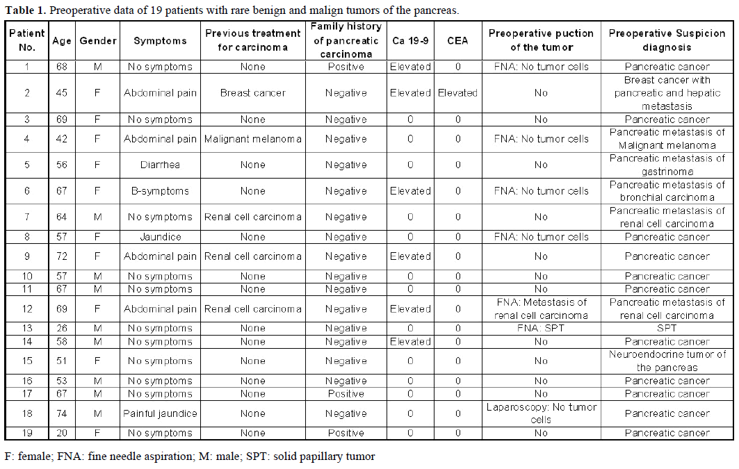
After completion of staging diagnostics, metastatic disease to the pancreas was assumed in 6 patients (31.6%) who had been previously treated for renal carcinoma (n=3), breast cancer (n=1), melanoma (n=1) and in one patient with duodenal gastrinoma. In one patient a solid pseudopapillary tumor of the pancreas was suspected and in one patient a neuroendocrine tumor of the pancreas. The majority of patients (n=11; 57.9%) presented with an intrapancreatic mass that was suspicious for pancreatic cancer (Table 1).
Preoperative imaging in all patients revealed a solid intrapancreatic mass, thus malignancy could not be excluded. Before referral to our clinic, in 6 patients (31.6%) endoscopic ultrasound and fine needle aspiration of the tumor and in one patient (5.3%) laparoscopic biopsy were performed at other medical institutions. In only two cases (by fine needle aspiration; 10.5%) atypical cells could be separated that preoperatively lead to the diagnoses of a solid pseudopapillary tumor of the pancreas and a pancreatic metastasis of a renal cell carcinoma. Thus, before surgery a correct preoperative diagnosis of the tumor was only made in two patients (10.5%). In the remaining 17 patients (89.5%) operation was necessary to affirm the diagnosis and to supply the patient the optimal therapy (Table 1).
Surgical intervention was carried out with different intentions. Ten patients (52.6%) were resected with curative intent. In 4 patients (21.1%) palliative tumor debulking and in one patient (5.3%) bypass operation were performed. Solely sampling of biopsies for histologic confirmation of tumor entity was done in 4 patients (21.1%) (Table 2).

Postoperative histological examination of the resected specimen or taken biopsies revealed pancreatic metastases of extrapancreatic malignomas (Figure 1) in 8 patients (renal cell carcinoma, n=3; melanoma, n=2; duodenal gastrinoma, n=1; breast cancer, n=1; retroperitoneal liposarcoma, n=1). In 10 patients (52.6%) the following rare benign tumors were detected: solid pseudopapillary tumor of the pancreas (n=3) (Figure 2), mature teratoma of the pancreas (n=2) (Figure 3), capillary hemangioma of the pancreas (n=1) (Figure 4), intrapancreatic accessory spleen (n=1) (Figure 5), lymphoepithelial cyst of the pancreas (n=1) (Figure 6), hamartoma of the pancreas (n=1) (Figure 7), and pancreatic sarcoidosis (n=1). In one patient an advanced yolk sac tumor of the pancreas with peritoneal carcinosis was diagnosed (Table 2).
Figure 1. Intrapancreatic metastasis of a renal cell carcinoma
(arrow). a. Preoperative CT scan. b. Histology of a pancreatic
metastasis of a renal clear cell carcinoma (G2); tumor cell clusters
(arrow), pancreatic parenchyma with chronic inflammation on the
right (H&E-staining).
Figure 2. Solid pseudopapillary tumors of the pancreas (arrow). a. Preoperative CT scan. b. Intraoperative photograph. c. Histology of a
solid pseudopapillary tumor of the pancreas with characteristic
pseudopapillary morphology of monomorphous discohesive tumor
cells on the left (arrow) and intratumoral foamy macrophages
surrounding cholesterol clefts on the lower right (H&E-staining).
Figure 3. Teratoma of the pancreas. Histology of mature teratoma of
the pancreas with predominant squamous epithelium as well as
lymphatic tissue and connective tissue, normal pancreatic tissue on
the lower right (H&E-staining).
Figure 4. Capillary hemangioma of the pancreas (arrow). a. Intraoperative photograph. b. Macroscopic photograph. c. Histology
of a capillary hemangioma of the pancreas with central regressive
changes (arrow) (H&E-staining).
Figure 5. Intrapancreatic accessory spleen (arrow). a. Macroscopic
photograph. b. Histology of an accessory intrapancreatic spleen with
red and white pulpa (arrow) with a fibrous capsule next to normal
pancreatic tissue (H&E-staining).
Figure 6. Lymphoepithelial cyst of the pancreas (arrow). a. Preoperative MRI scan. b. Macroscopic photograph. c. Histology of
a lymphoepithelial cyst of the pancreas. A cystic space lined with flat
squamous epithelium and hyperplastic lymphatic follicles, on the
right pancreatic tissue with chronic inflammatory changes (H&Estaining).
Figure 7. Histology of a hamartoma of the pancreas. A
circumscribed area of fibrous tissue with embedded acinar structures
and small ductules and adipous tissues in the pancreatic head (H&Estaining).
Postoperative complications occurred in 8 patients (42.1%). One patient needed operative revision following pancreatic grade B fistula [7]. Three more patients were treated by minimal invasive interventions following pancreatic grade B fistula, intra-abdominal abscess and iatrogenic lesion of the ureter. Wound infection, pneumonia and renal failure regressed under conservative treatment. Two patients died during the postoperative course (10.5%). One patient with pancreatic metastasis of a renal cell carcinoma died following postoperative bleeding from the splenic artery. Another patient with pancreatic metastasis of a melanoma died of tumor progression (Table 2).
DISCUSSION
Advancement of imaging techniques and the improved awareness of clinical and pathological features of pancreatic neoplasms increasingly lead to the detection of rare solid intrapancreatic neoplasms that are difficult to differentiate from primary pancreatic ductal adenocarcinoma or neuroendocrine tumors.
From 2004 to 2010 we operated 19 patients with rare pancreatic neoplasms. After extended preoperative diagnostics in the majority of patients (n=11) pancreatic cancer was suspected. Two of these patients presented with elevated tumor markers (Table 1), two other patients with jaundice and three patients reported a positive family history of pancreatic carcinoma. At other medical institutions endoscopic ultrasound and fine needle biopsy were done in two patients and diagnostic laparoscopy was done in one patient without any histological evidence. At our institution, intraoperative examination of frozen sections of the resected specimens and postoperative histological investigations excluded pancreatic cancer and detected in 10 patients the below described rare benign pancreatic tumors.
In a 68-year-old male patient without any medical history of sarcoidosis, pancreatic sarcoidosis was the cause of a mass in the head of the pancreas accompanied by an elevation of the tumor marker CA 19-9 (199 U/mL; reference range: 0-37 U/mL). Pancreatic sarcoidosis is extremely uncommon [8, 9]. The first case was described on autopsy in 1937 [10]. Comparable to our case, the patients present with all the signs and symptoms of a pancreatic malignancy, which was confirmed on a CT scan. The CA 19-9 level is also confirmatory of the suspected diagnosis [10]. Comparable to our case, the disease most often presents as a pancreatic head mass. The preoperative diagnosis of this entity is a clinical challenge, and surgical intervention is usually needed to make a definitive diagnosis [11].
In two male patients (57 and 58 years of age) an asymptomatic mass of 4 cm in the tail of the pancreas was identified as mature teratoma and distal pancreatectomy was performed. In one patient the tumor marker CA 19-9 was elevated (66 U/mL). Teratoma of the pancreas were first described in 1918 by Kerr [12]. They can be classified as benign, welldifferentiated lesions, which are solid or cystic, and solid malignant undifferentiated tumors, named, respectively, mature and immature teratomas [13]. Surgical therapy is the only way of guaranteeing definitive resolution [14, 15, 16]. Even though ultrasound, CT and MRI may be helpful, there are no pathognomonic data for their preoperative recognition [17].
A 74-year-old male patient was hospitalized following painful jaundice. MRI and CT showed a double duct sign and a mass of 8 cm diameter in the pancreatic head. Stenting of the common bile duct and diagnostic laparoscopy for taking biopsies was performed at another hospital before. As the mass was suspicious for carcinoma and biopsies did not confirm the diagnosis, surgical exploration was done at our department. A large tumorous mass was found in the pancreatic head and peritumorous inflammation involved vessels, stomach and the rest of the pancreas thus total pancreatectomy was carried out. Histological examination of the resected specimen revealed a benign lymphoepithelial cyst of the pancreas. Lymphoepithelial cyst is a rare benign lesion which was described for the first time in 1987 by Truong et al. [18]. Histologically, the lesion has a complex content consisting of keratinous material and a wall lined with mature squamous epithelium surrounded by dense lymphoid tissue [19]. The most common symptoms are abdominal pain, nausea and vomiting, anorexia and weight loss, but many patients are asymptomatic, coming to the surgeon’s attention as incidental radiological finding [19]. Lymphoepithelial cyst may appear either multilocular (60%) or unilocular (40%) as described in our case. The etiopathogenesis as well as histogenesis of lymphoepithelial cyst remain unclear. They have been described in other locations which are associated with autoimmune diseases and states of immunological depression, frequently [20]. Imaging may be not specific and the radiological appearance of these lesions differs. Surgical resection should still be considered the standard therapy, in suitable patients, to exclude malignancy [21]. The prognosis is fairly good. There has never been a report of local recurrence after operative resection.
Another 53-year-old male patient underwent extirpation of a 8 cm diameter asymptomatic hemangioma of the pancreatic head. Pancreatic hemangiomas are extremely uncommon benign pancreatic vascular neoplasms [22]. In contrast to infantile hemangioma - that mostly presents before 6 months of age, grows rapidly, and then regresses spontaneously over several months [23] - adult pancreatic hemangiomas do not regress and reveal a risk of bleeding. As in our case they may not contrast enhance on arterial phase CT imaging [24], thus pancreatic cancer was suspected.
An intrapancreatic accessory spleen was found in a 67- year-old male patient. A mass of 1.5 cm diameter in the pancreatic tail was detected in a routine check-up. Due to a family history of pancreatic cancer (mother and sister) surgical exploration and distal pancreatectomy were done. Autopsy studies suggest that in 80% the accessory spleen is located at or near the splenic hilum. The second most common site is the pancreatic tail [25, 26]. As in our patient, most often an intrapancreatic accessory spleen is small with a diameter of less than 2 cm [27]. Generally an intrapancreatic accessory spleen does not usually require treatment. Unfortunately, current CT, MRI, and ultrasound technologies do not necessarily distinguish between splenic tissue and pancreatic neuroendocrine neoplasms. Only nuclear medicine examinations/scintigraphy may confirm the diagnosis [28]. Our decision for surgical exploration due to a family history of pancreatic cancer is in line with the statement of Meiler et al. [29] that intrapancreatic accessory spleen is a rare cause of unnecessary laparotomy, but the absence of reliable diagnostics for this entity makes histologic ascertainment of a benign tumor indispensable.
An asymptomatic mass of 5 cm diameter in the processus uncinatus of the pancreas, enlarged peritumoral lymph nodes and a hypodense liver structure (segment VII) suggested a malignant pancreatic tumor in a 67-year-old male patient. MRI showed a stenosis of the pancreatic duct and PET-CT did not show any significantly increased activity. Total pancreatectomy was performed due to a soft fatty pancreatic tissue. Histopathological examination confirmed the diagnosis of a pancreatic hamartoma. A hamartoma is a mass composed of an excess of differentiated cells or mixture of cell types that are normally present in the organ where the mass is found. It may be regarded as a malformation rather than a neoplasm [30]. As in our case nearly all hamartoma arise in the head of the pancreas and tend to affect mostly males [31]. Surgical resection and histopathological examination are required to confirm the diagnosis [31].
In three patients (a 20-year-old female, a 51-year-old female, and a 26-year-old male) a solid pseudopapillary tumor of the pancreas was diagnosed. Solid pseudopapillary tumor (Franz tumor) accounts for less than 1% of all pancreatic tumors [32]. It is of lowgrade malignancy but can cause extensive local invasion [33]. Our patients did not show any symptom which is typical for solid pseudopapillary tumors, that are commonly detected incidentally on imaging studies for other reasons [34, 35]. In the male patient a diagnostic biopsy of the tumor, that was carried out before referral to our department, revealed the diagnosis before surgery. The 20-year-old female had a family history of pancreatic cancer (uncle, grandmother). CT and MRCP in all patients showed solid and cystic fractions of the tumor. Butte et al. [36] published data of 45 patients with solid pseudopapillary tumors. Their results demonstrated that solid pseudopapillary tumors primarily occur in young women. Only about 8.3% of all cases were reported in males [37, 38]. About 15% are known to present with metastasis or recurrence [39]. The only feature associated with malignant disease is tumor size (7.8 vs.4.2 cm) at presentation [36]. In our patients tumors had a size between 2 and 4.5 cm and were located at different sites of the organ (head, body, tail). Oncologic outcomes in patients who undergo surgical resection are excellent [32]. This is in line with our observations. Until July 2011, 10 to 29 months after operation (Whipple operation, distal pancreatectomy, local excision of the solid pseudopapillary tumor), all patients are in good health without relapse. Surgery including enucleation is typically curative in patients with localized disease and possibly in patients with limited metastasis [40, 41, 42]. No consensus exists on an effective systemic therapy or radiation [43].
In contrast to the above described rare benign intrapancreatic neoplasms, in a 69-year-old female patient, who presented with a mass in the body of the pancreas suspicious for pancreatic cancer, a malignant yolk sac tumor with peritoneal carcinosis and local infiltration of the stomach was diagnosed following surgical exploration. Yolk sac tumor also known as endodermal sinus tumor and Teilum tumor, is one type of germ cell neoplasm. Of all the genital tumors, yolk sac tumors are relatively uncommon and, unlike our case, they are mostly discovered in infants and adolescents (median age: 19 years) [44]. Yolk sac tumor is considered to be a highly malignant tumor that primarily occurs in the testis or ovary [45]. Ten to fifteen percent of these tumors may arise in a variety of midline extragonadal sites. Exceedingly rare sites such as liver, kidney, omentum, stomach, spinal cord and pancreas have been reported [45, 46, 47, 48]. The imaging findings were verified by the morphological observations of an encapsulated tumor with focal necrosis. However in our patient CT was without any pathologic findings. The tumor in the body of the pancreas with a diameter of 3 cm was only detected by endoscopic ultrasound. Since yolk sac tumor usually show high malignancy, the duration from the onset of symptoms to the admission is always short and, as in our patient, metastasis may already exist at the time of the patient’s admission. Surgical excision with combined adjuvant chemotherapy, as it was performed in our patient, is the treatment of choice. However, the prognosis is poor if there is metastasis [45].
Eight patients presented with metastatic tumors to the pancreas (3 renal cell carcinomas, 2 melanomas, 1 duodenal gastrinoma, 1 breast cancer, and 1 retroperitoneal liposarcoma). Metastatic tumors to the pancreas account for less than 2% of all pancreatic malignancies [49]. As with primary pancreatic cancer, early signs and symptoms of isolated pancreatic metastases are often nonspecific and subtle. Patients without symptoms at the time of diagnosis (43%) account for the largest group [49]. In our collective one patient was asymptomatic, four patients complained about abdominal pain, one patient presented with jaundice, one patient with B symptoms (fever, night sweats, weight loss) and the patient with metastatic gastrinoma had diarrhea. Most patients with a pancreatic secondary tumor are not candidates for resection because they have widespread disease. In our collective in three patients (two patients with renal cell carcinoma and one patient with breast cancer) surgical exploration showed widespread disease and resection was not possible. For improvement of life quality in one of these patients a palliative double bypass was created. Comparable to our observations most pancreatic metastases are referable to renal cell carcinoma [50]. However, metastases from primary lung, breast, colon, skin (melanoma), and sarcoma tumors also involve the pancreas [3]. In accordance with the current literature, metastasectomy with curative intent has become standard practice for the management of metastatic lesions to the liver, lung, and brain from several tumors, with a clear survival benefit [51]. The effectiveness of pancreatic metastasectomy is dependent on the tumor biology of the primary cancer. Renal cell carcinoma is associated with the best outcome, whereas lung cancer predicts the worst outcome [51, 52]. Patients with renal cell cancer may present with metastases to the pancreas many years after the initial diagnosis, emphasizing the need for lifelong surveillance. They can have a good long-term prognosis after surgical resection, with a 5- year survival rate up to 88% in some series [35, 53, 54, 55]. The results of chemotherapy in melanoma are generally disappointing. Presently, surgical resection appears to be the only potentially curative treatment option. However, all metastases have to be excised if surgery is to offer any survival benefit [56]. The survival values after resection of sarcomas are substantially lower than what has been reported for either lung or liver metastasectomies [51]. Nevertheless, pancreatic surgery is considered to be one of the most technically demanding and challenging surgical disciplines and it has been associated with a high rate of mortality and morbidity, which has declined for the past 3 decades. In our collective survival rates were 2 months for renal cell carcinoma, 3 months for melanoma, 4 months for breast cancer, 17 months for sarcoma and 62 months for gastrinoma.
Two patients died in the postoperative course: a 67- year-old woman with melanoma died following tumor progress after palliative double bypass and a 64-yearold male patient underwent bleeding of the splenic artery and died after reoperation. One patient had a complicated postoperative course after total pancreatectomy with pneumonia and renal failure. Three patients underwent curative surgery (2 Whipple operations, 1 distal pancreatectomy) and one patient underwent palliative exploration without any complications. Despite the above mentioned morbidity and mortality rates the potential benefit of metastasectomy is documented and it can improve quality of life and survival time being the only chance of cure in selected patients [57, 58].
CONCLUSION
With regard to the presented collective of patients with rare benign and malignant tumors of the pancreas, we conclude, that sometimes even extensive imaging cannot certify the nature of an intrapancreatic tumor. Similar to pancreatic cancer, benign and malignant tumors of the pancreas, intrapancreatic metastasis, pancreatic malformations and abnormalities present as solid masses of the pancreas. Thus, they constitute a differential diagnosis to pancreatic adenocarcinoma. Elevated CA 19-9 levels or a positive family history of pancreatic cancer make malignancy even more likely. Percutaneous or endoscopic ultrasound-guided fine needle aspiration biopsy sometimes can help to distinguish a benign neoplasm from malignant pancreatic tumor [59]. However, there are several reports available on seeding of the needle tract by neoplastic cells and complications such as bleeding, pancreatic fistula and biliary fistula [60]. Steady technical improvement in pancreatic surgery and advances in perioperative supportive care has significantly ameliorated postoperative outcome [61]. Thus, radical resection, which remains the only curative treatment option in adenocarcinoma of the pancreas, can be performed with low mortality and morbidity rates in specialized centers. We conclude that in cases of doubtful dignity, operative exploration, biopsy and when indicated resection of the lesion should be performed. Also for solitary metastases to the pancreas, solid pseudopapillary tumor, yolk sac tumor and symptomatic benign neoplasms of the pancreas there is no satisfactory non-surgical treatment, currently.
Note
Sabine Kersting and Monika S Janot equally contributed to this study
Conflicts of interest
The authors have no potential conflicts of interest
References
- Peters K, Kloppel G. [Undifferentiated pancreatic carcinomas.Leap into chaos]. Pathologe 2005;26:18-21.
- Carpelan-Holmstrom M, Nordling S, Pukkala E, Sankila R,Luttges J, Kloppel G et al. Does anyone survive pancreatic ductaladenocarcinoma? A nationwide study re-evaluating the data of theFinnish Cancer Registry. Gut 2005;54:385-387.
- Adsay NV, Basturk O, Klimstra DS, Kloppel G. Pancreaticpseudotumors: non-neoplastic solid lesions of the pancreas thatclinically mimic pancreas cancer. SeminDiagnPathol 2004;21:260-267.
- Mortenson MM, Katz MH, Tamm EP. Current diagnosis andmanagement of unusual pancreatic tumors. Am J Surg 2008;196:100-113.
- Roland CF, van Heerden JA. Nonpancreatic primary tumorswith metastasis to the pancreas. SurgGynecolObstet 1989;168:345-347.
- Z'Graggen K, Fernandez-del Castillo C, Rattner DW, Sigala H,Warshaw AL. Metastases to the pancreas and their surgicalextirpation. Arch Surg 1998;133:413-417; discussion 418-419.
- Pratt WB, Maithel SK, Vanounou T, Huang ZS, Callery MP,Vollmer CM. Clinical and economic validation of the InternationalStudy Group of Pancreatic Fistula (ISGPF) classification scheme.Ann Surg 2007;245:443-451.
- Baroni RH, Pedrosa I, Tavernaraki E, Goldsmith J, Rofsky NM.Pancreatic sarcoidosis: MRI features. J MagnReson Imaging2004;20:889-893.
- Fong ZV, Wong J, Maley WR, Sandorfi N, Winter J M, KoniarisLG et al. Sarcoid-Reaction Mimicking Metastatic MalignantHepatopancreatobiliary Tumors: Report of Two Cases and Review ofthe Literature. J GastrointestSurg 2012.
- Wijkstrom M, Bechara RI, Sarmiento JM. A rare nonmalignantmass of the pancreas: case report and review of pancreaticsarcoidosis. Am Surg 2010;76:79-84.
- Caceres M, Sabbaghian MS, Braud R, Wilks S, Boyle M.Pancreatic sarcoidosis: unusual presentation resembling aperiampullary malignancy. Curr Surg 2006;63:179-185.
- Kerr AA. Cysts and pseudocysts of the pancreas. SurgGynecolObstet 1918;27:40.
- Koomalsingh KJ, Fazylov R, Chorost MI, Horovitz J. Cysticteratoma of the pancreas: presentation, evaluation and management.Jop 2006;7:643-646.
- Degrate L, Misani M, Mauri G, Garancini M, Maternini M,Moltrasio F et al. Mature cystic teratoma of the pancreas. Case reportand review of the literature of a rare pancreatic cystic lesion. Jop2012;13:66-72.
- Iacono C, Zamboni G, Di Marcello R, Zicari M, Maran M,Montresor E et al. Dermoid cyst of the head of the pancreas area. IntJ Pancreatol 1993;14:269-273.
- Scheele J, Barth TF, Wittau M, Juchems M, Henne-Bruns D,Kornmann M. Cystic teratoma of the pancreas. Anticancer research2012;32:1075-1080.
- Adsay NV, Hasteh F, Cheng JD, Klimstra DS. Squamous-linedcysts of the pancreas: lymphoepithelial cysts, dermoid cysts(teratomas), and accessory-splenic epidermoid cysts. SeminDiagnPathol 2000;17:56-65.
- Truong LD, Rangdaeng S, Jordan PH, Jr. Lymphoepithelial cystof the pancreas. Am J SurgPathol 1987;11:899-903.
- Matrone A, Russo M, Mollica C, Lombardi D, Lombardi G,Maurea S et al. Lymphoepithelial pancreatic cyst: an atypical benignpancreatic mass presenting with a "cheerios-like" appearance. Jop2010;11:170-172.
- Jian B, Kimbrell HZ, Sepulveda A, Yu G. Lymphoepithelialcysts of the pancreas: endosonography-guided fine needle aspiration.DiagnCytopathol 2008;36:662-665.
- Tucci G, Muzi MG, Nigro C, Cadeddu F, Amabile D, ServadeiFet al. Dermoid cyst of the pancreas: presentation and management.World J SurgOncol 2007;5:85.
- England RJ, Woodley H, Cullinane C, McClean P, Walker J,Stringer M D. Pediatric pancreatic hemangioma: a case report andliterature review. Jop 2006;7:496-501.
- Tebboune N, Lazure T, Fabre M, Pariente D. Pancreatichaemangioma in infancy: the place of radiology. PediatrRadiol2003;33:621-623.
- Mundinger GS, Gust S, Micchelli ST, Fishman EK, Hruban RH,Wolfgang CL. Adult pancreatic hemangioma: case report andliterature review. Gastroenterol Res Pract 2009;2009:839730.
- Halpert B, Gyorkey F. Lesions observed in accessory spleens of311 patients. Am J ClinPathol 1959;32:165-168.
- Rodriguez E, Netto G, Li QK. Intrapancreatic accessory spleen:A case report and review of literature. Diagnostic cytopathology2012.
- Guo W, Han W, Liu J, Jin L, Li JS, Zhang ZT et al.Intrapancreatic accessory spleen: a case report and review of theliterature. World J Gastroenterol 2009;15:1141-1143.
- Spencer LA, Spizarny DL, Williams TR. Imaging features ofintrapancreatic accessory spleen. Br J Radiol 2010;83:668-673.
- Meiler R, Dietl KH, Novak K, Patzel C. Intrapancreaticaccessory spleen. IntSurg 2010;95:183-187.
- Longnecker DS. Endocrine Tumors of the Pancreas inTransgenic Mice. Comp Med 2004;54:21-22.
- McFaul CD, Vitone LJ, Campbell F, Azadeh B, Hughes ML,Garvey CJ et al. Pancreatic hamartoma. Pancreatology 2004;4:533-537; discussion 537-538.
- Matos JM, Grutzmann R, Agaram NP, Saeger HD, Kumar HR,Lillemoe KD et al. Solid pseudopapillary neoplasms of the pancreas:a multi-institutional study of 21 patients. J Surg Res 2009;157:e137-142.
- Campanile M, Nicolas A, LeBel S, Delarue A, Guys JM, deLagausie P. Frantz's tumor: is mutilating surgery always justified inyoung patients? SurgOncol 2011;20:121-125.
- Chakhachiro ZI, Zaatari G. Solid-pseudopapillary neoplasm: apancreatic enigma. Arch Pathol Lab Med 2009;133:1989-1993.
- Zerbi A, Ortolano E, Balzano G, Borri A, Beneduce AA, DiCarlo V. Pancreatic metastasis from renal cell carcinoma: whichpatients benefit from surgical resection? Ann SurgOncol2008;15:1161-1168.
- Butte JM, Brennan MF, Gonen M, Tang LH, D'Angelica MI,Fong Y et al. Solid pseudopapillary tumors of the pancreas. Clinicalfeatures, surgical outcomes, and long-term survival in 45 consecutivepatients from a single center. J GastrointestSurg 2011;15:350-357.
- Martin RC, Klimstra DS, Brennan MF, Conlon KC. Solidpseudopapillarytumor of the pancreas: a surgical enigma? Ann SurgOncol 2002;9:35-40.
- Tien YW, Ser KH, Hu RH, LeeCY, Jeng YM, Lee PH et al.Solid pseudopapillary neoplasms of the pancreas: is there apathologic basis for the observed gender differences in incidence?Surgery 2005;137:591-596.
- Tang LH, Aydin H, Brennan MF, Klimstra DS. Clinicallyaggressive solid pseudopapillary tumors of the pancreas: a report oftwo cases with components of undifferentiated carcinoma and acomparative clinicopathologic analysis of 34 conventional cases. AmJ SurgPathol 2005;29:512-519.
- Lee JS, Han HJ, Choi SB, Jung CW, Song TJ, Choi SY. Surgicaloutcomes of solid pseudopapillary neoplasm of the pancreas: a singleinstitution's experience for the last ten years. The American surgeon2012;78:216-219.
- Nakagohri T, Kinoshita T, Konishi M, Takahashi S, Gotohda N.Surgical outcome of solid pseudopapillary tumor of the pancreas. JHepatobiliaryPancreatSurg 2008;15:318-321.
- Reddy S, Cameron JL, Scudiere J, Hruban RH, Fishman EK,Ahuja N et al. Surgical management of solid-pseudopapillaryneoplasms of the pancreas (Franz or Hamoudi tumors): a largesingle-institutional series. J Am CollSurg 2009;208:950-957;discussion 957-959.
- Reddy S, Wolfgang CL. Solid pseudopapillary neoplasms of thepancreas. AdvSurg 2009;43:269-282.
- Razzi S, Luisi S, Gabbanini M, Lazzeri L, Mazzini M, PetragliaF. Yolk sac tumor in a young girl: a case report. GynecolEndocrinol2005;20:334-335.
- Zhang B, Gao S, Chen Y, Wu Y. Primary yolk sac tumor arisingin the pancreas with hepatic metastasis: a case report. Korean JRadiol 2010;11:472-475.
- Guzel A, Tatli M, Belen D, Seckin H. Spinal cord compressionof primary extragonadal giant yolk sac tumor. Spinal Cord2007;45:254-257.
- Kumar Y, Bhatia A, Kumar V, Vaiphei K. Intrarenal pure yolksac tumor: an extremely rare entity. Int J SurgPathol 2007;15:204-206.
- Park NH, Ryu SY, Park IA, Kang SB, Lee HP. Primaryendodermal sinus tumor of the omentum. GynecolOncol1999;72:427-430.
- Mourra N, Arrive L, Balladur P, Flejou JF, Tiret E, Paye F. .Isolated metastatic tumors to the pancreas: Hopital St-Antoineexperience. Pancreas 2010;39:5 77-580.
- Thompson LD, Heffess CS. Renal cell carcinoma to the pancreasin surgical pathology material. Cancer 2000;89:1076-1088.
- Reddy S, Wolfgang CL. The role of surgery in the managementof isolated metastases to the pancreas. Lancet Oncol 2009;10:287-293.
- Sperti C, Pasquali C, Liessi G, Pinciroli L, Decet G, PedrazzoliS. Pancreatic resection for metastatic tumors to the pancreas. J SurgOncol 2003;83:161-166; discussion 166.
- Hiotis SP, Klimstra DS, Conlon KC, Brennan MF. Results afterpancreatic resection for metastatic lesions. Ann SurgOncol2002;9:675-679.
- Law CH, Wei AC, Hanna SS, Al-Zahrani M, Taylor BR, GreigPD. Pancreatic resection for metastatic renal cell carcinoma:presentation, treatment, and outcome. Ann SurgOncol 2003;10:922-926.
- Ballarin R, Spaggiari M, Cautero N, De Ruvo N, Montalti R,Longo C et al. Pancreatic metastases from renal cell carcinoma: thestate of the art. World J Gastroenterol 2011;17:4747-4756.
- Nikfarjam M, Evans P, Christophi C. Pancreatic resection formetastatic melanoma. HPB (Oxford) 2003;5:174-179.
- Reddy S, Edil BH, Cameron JL, Pawlik TM, Herman J M,Gilson MM et al. Pancreatic resection of isolated metastases fromnonpancreatic primary cancers. Ann SurgOncol 2008;15:3199-3206.
- Sweeney AD, Wu MF, Hilsenbeck SG, Brunicardi FC, FisherWE. Value of pancreatic resection for cancer metastatic to thepancreas. J Surg Res 2009;156:189-198.
- Layfield LJ, Hirschowitz SL, Adler DG. Metastatic disease tothe pancreas documented by endoscopic ultrasound guided fineneedleaspiration: a seven-year experience. Diagnostic cytopathology2012;40:228-233.
- Pettinato G, Di Vizio D, Manivel JC, Pambuccian SE, Somma P,Insabato L et al. Solid-pseudopapillary tumor of the pancreas: aneoplasm with distinct and highly characteristic cytological features.DiagnCytopathol 2002;27:325-334.
- Bassi C, Dervenis C, Butturini G, Fingerhut A, Yeo C, Izbicki Jet al. Postoperative pancreatic fistula: an international study group(ISGPF) definition. Surgery 2005;138:8-13.


